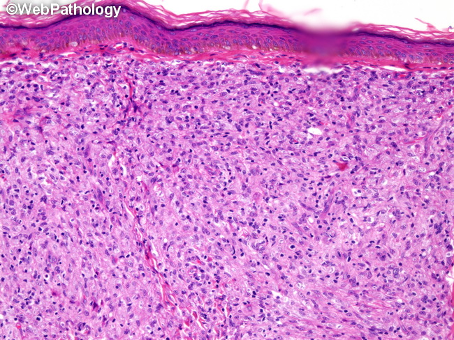
Leprosy Skin Histology
It is not a test to diagnose leprosy.
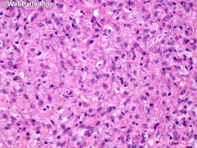
Leprosy skin histology. Clofazimine is added for the multibacillary having numerous bacilli form of leprosy. Macrophages may be distended with large groups of leprosy bacilli globi. Useful for multibacillary leprosy only.
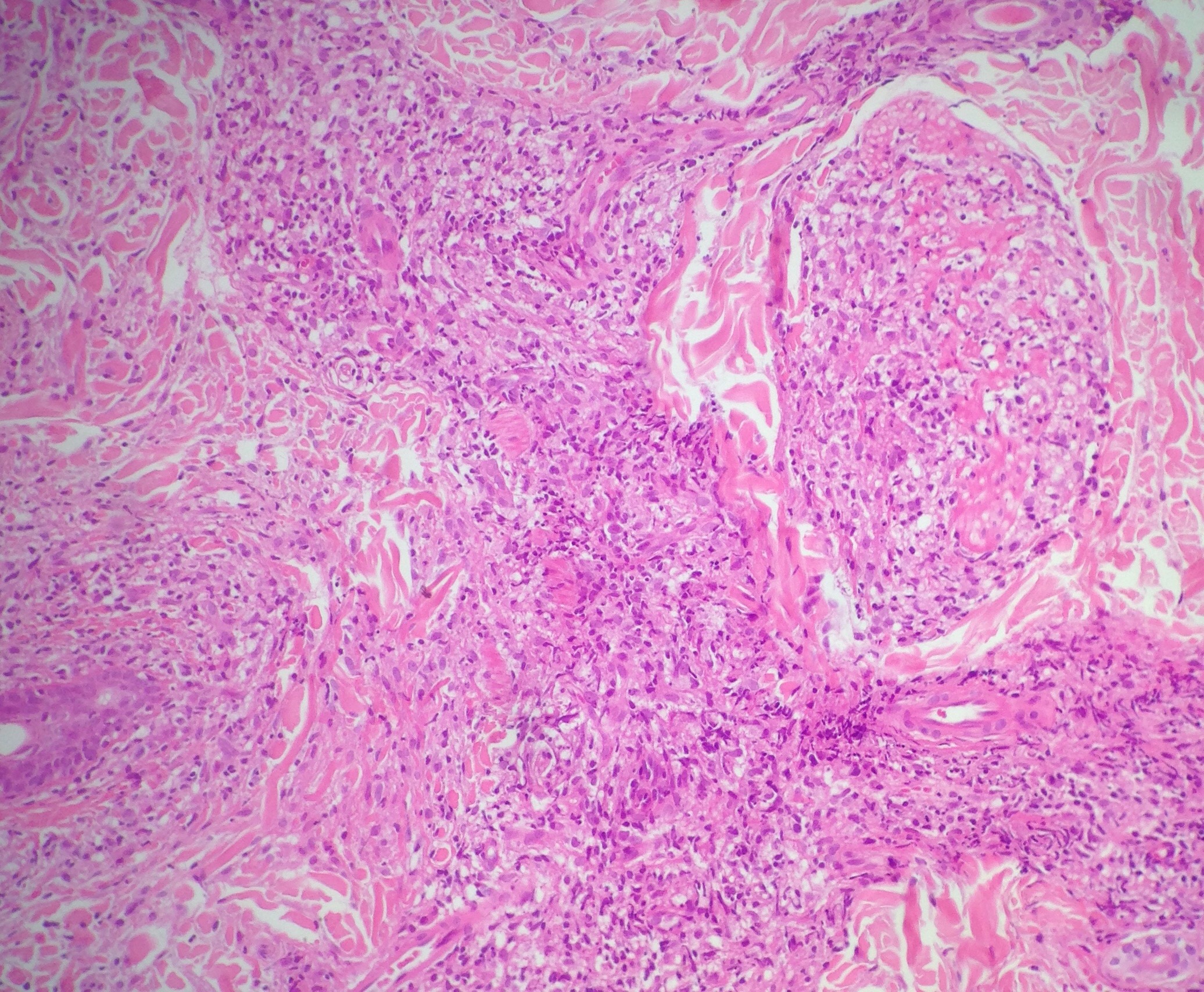
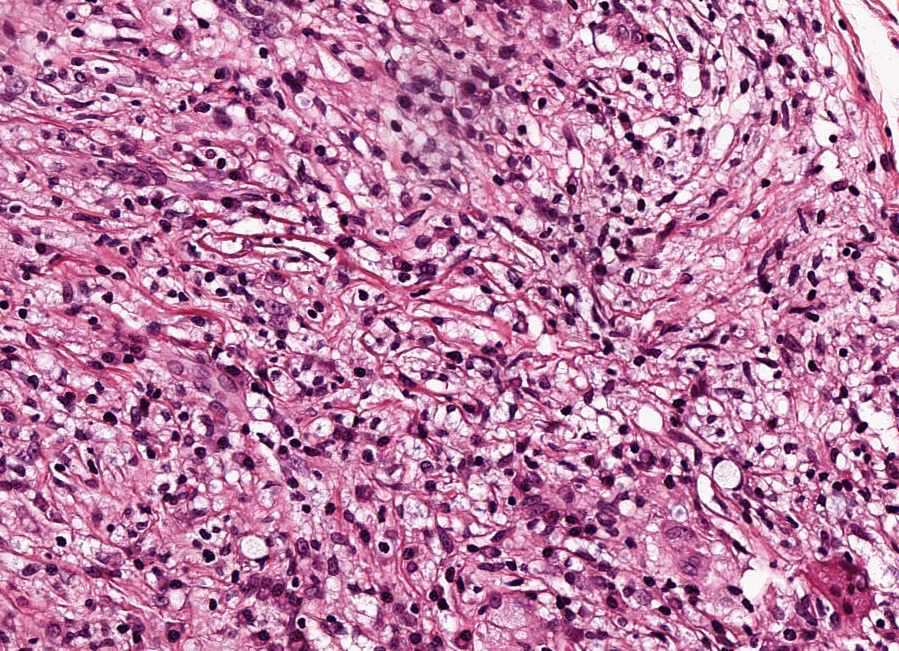
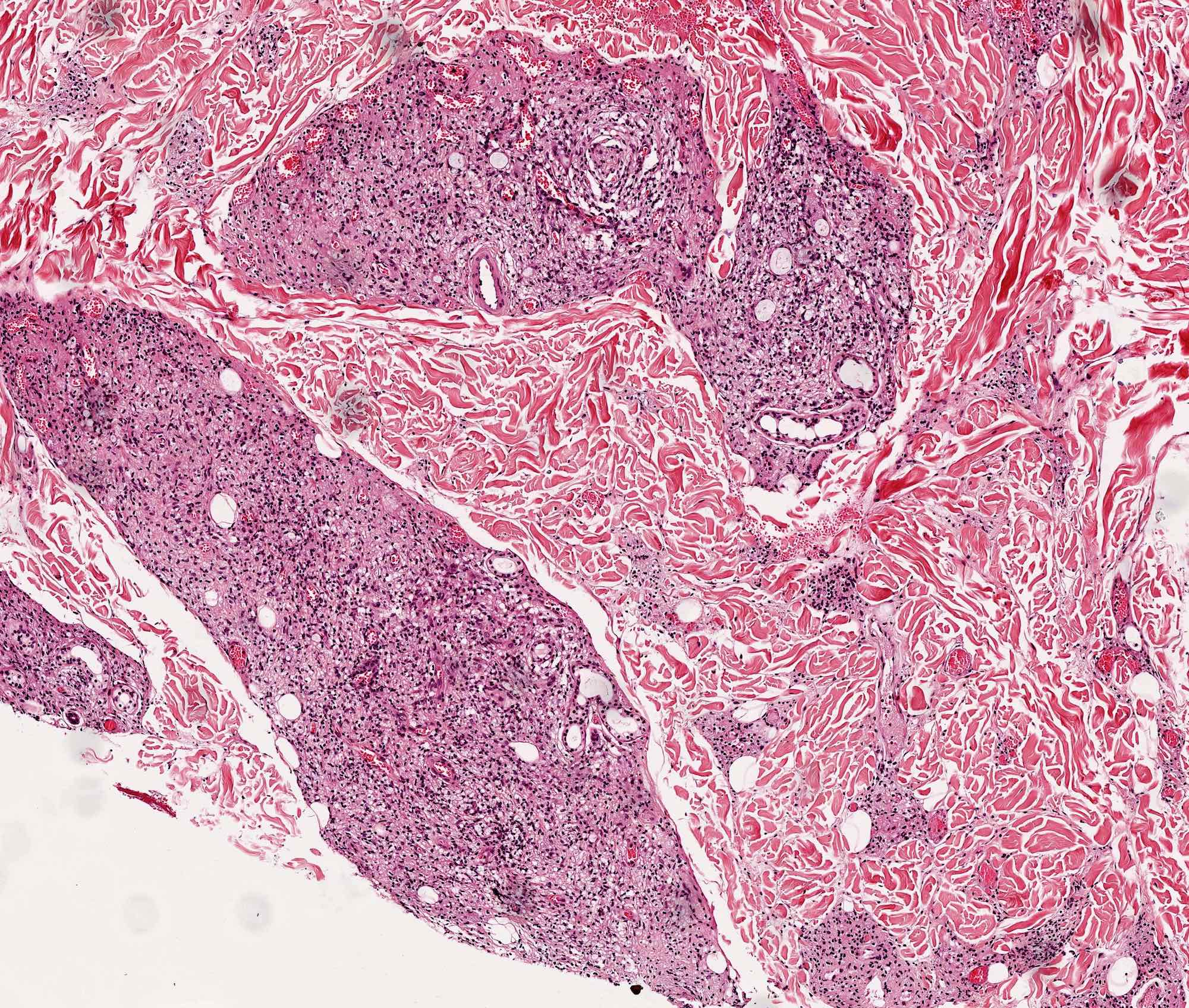
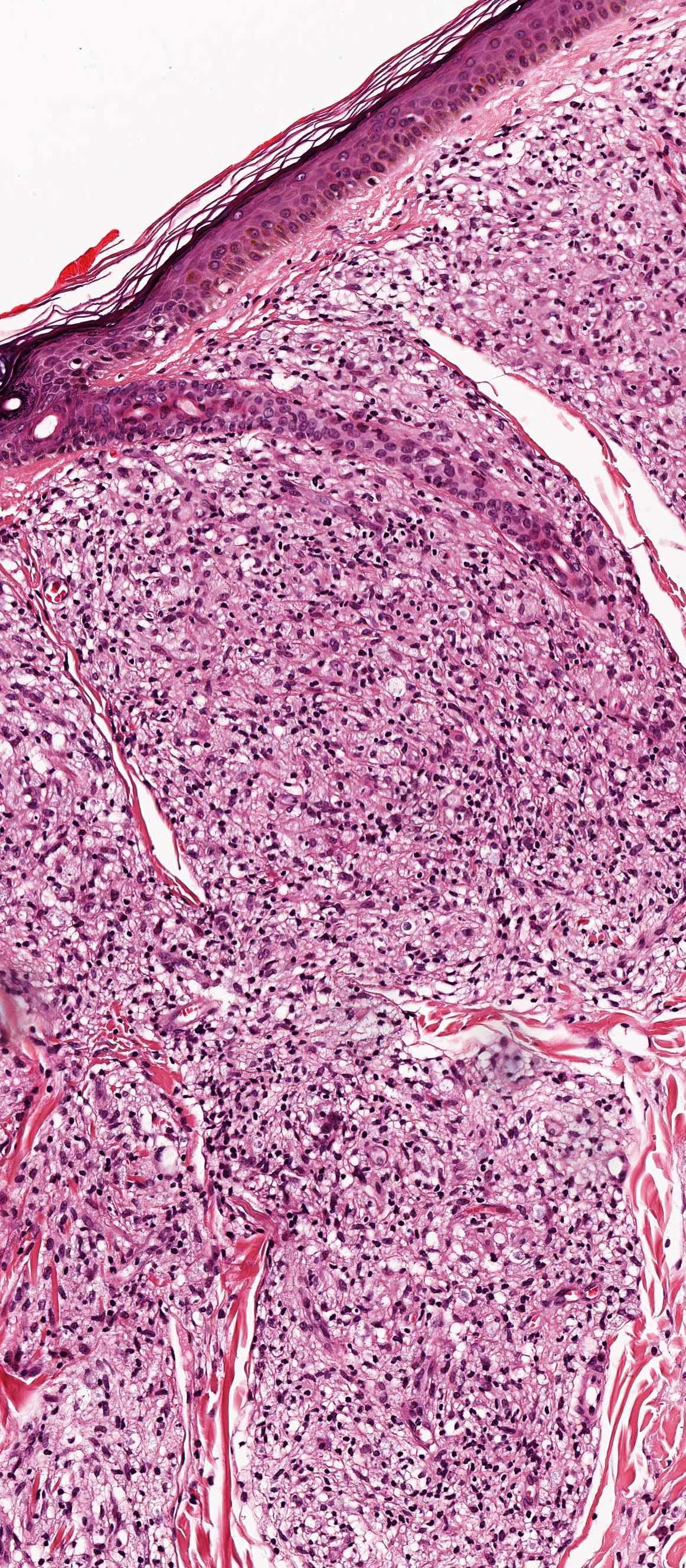
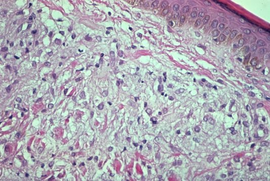
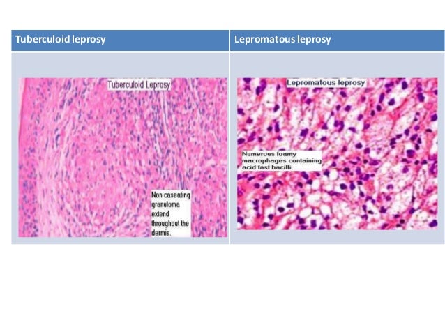
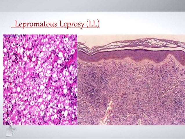
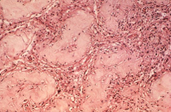
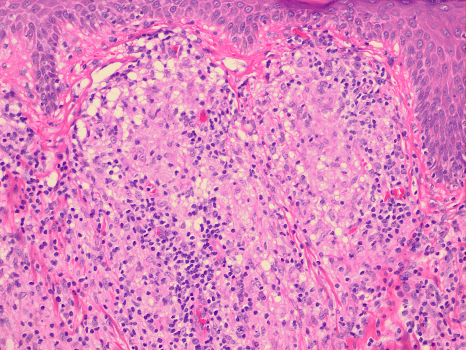
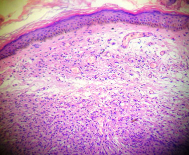
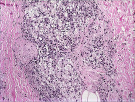
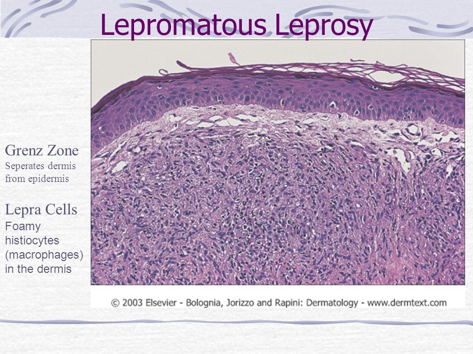
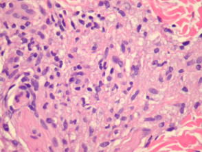
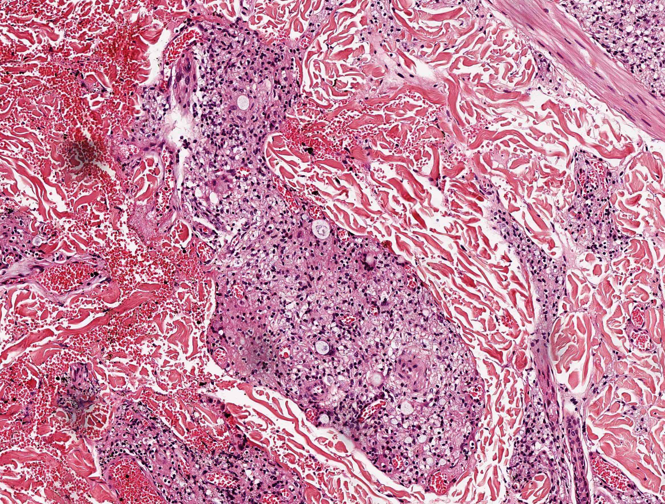
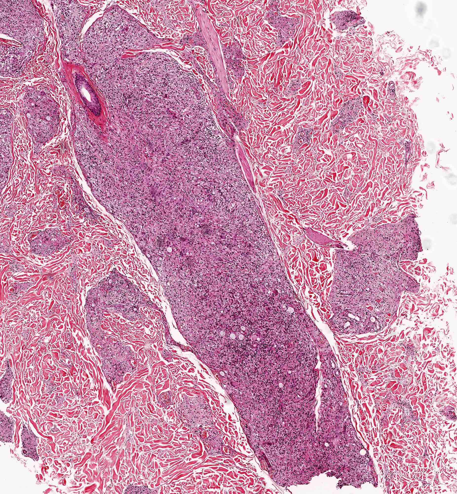
Lepromin skin test this is a test used to determine what type of leprosy a person has. It is similar to tuberculin test done for tb. Macrophages virchow cells lepra are found in poorly circumscribed masses in the dermis with few no lymphocytes.
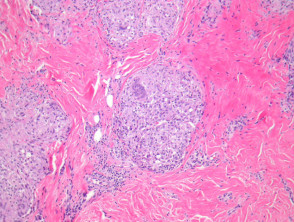
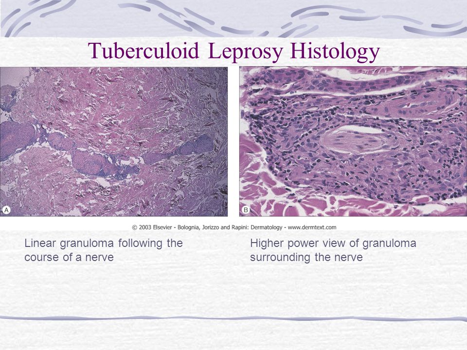
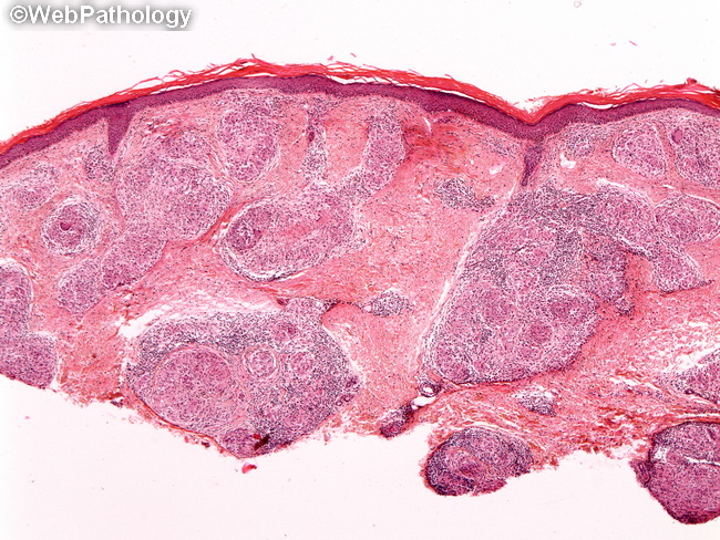
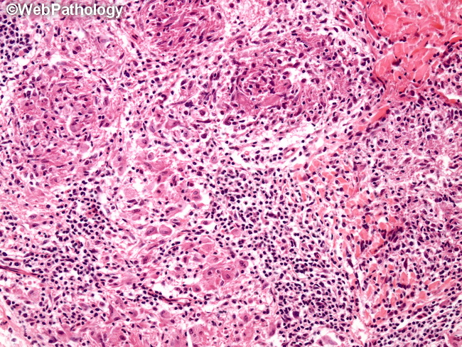
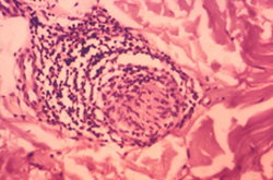
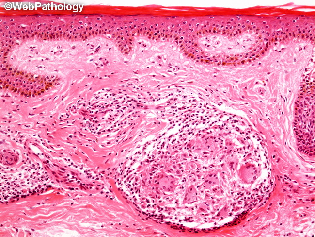
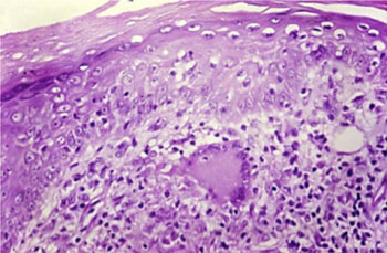
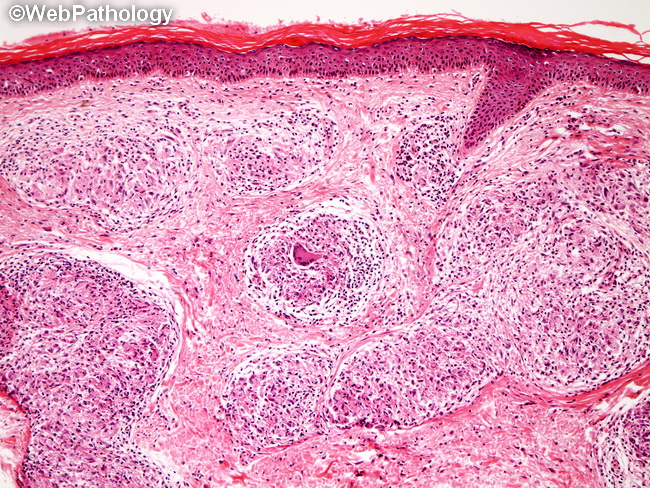
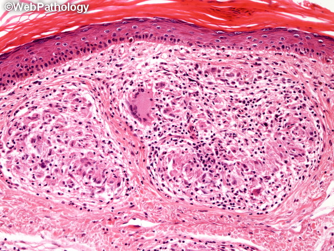
According to world health organization who leprosy is divided into two groups paucibacillary tt and tb and multibacillary bb bl and ll. No satisfactorily sensitive specific blood or skin tests are available for diagnosis at this time. In tuberculoid and borderline tuberculoid leprosy epithelioid noncaseating granulomas predominate and acid fast bacilli afb are absent or only rarely present.
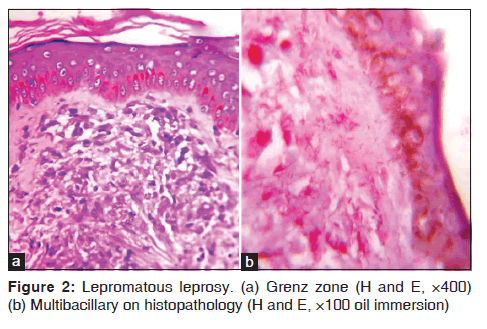
Leprosy has very characteristic clinical features but diagnosis should be confirmed by one of the following investigations. The macrophages have an abundant pink to pale cytoplasm which corresponds to numerous intracellular parasitized organisms figure 2. Curative treatment for the indeterminate stage of leprosy includes rifampicin and dapsone.
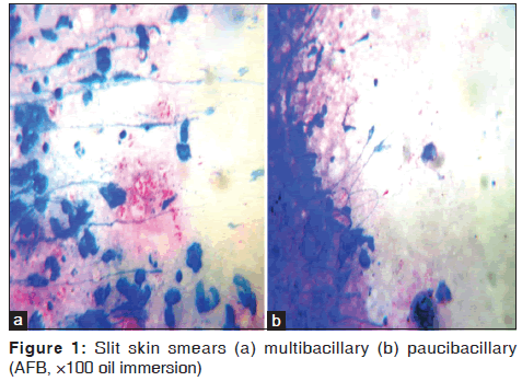
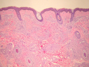
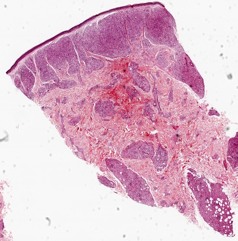
The classical clinical pattern of leprosy involves the skin nerves and nasal and oropharyngeal mucosa. Histology of leprosy lepromatous leprosy underneath a normal epidermis and grenz zone there are sheets or clusters of macrophages figure 1. Skin slit smear a small slit is made using a sharp blade over the skin of the earlobe forehead or lesional skin then a smear is made by scraping the exposed dermis onto a glass slide and examining for acid fast bacilli under microscopy.
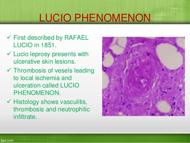
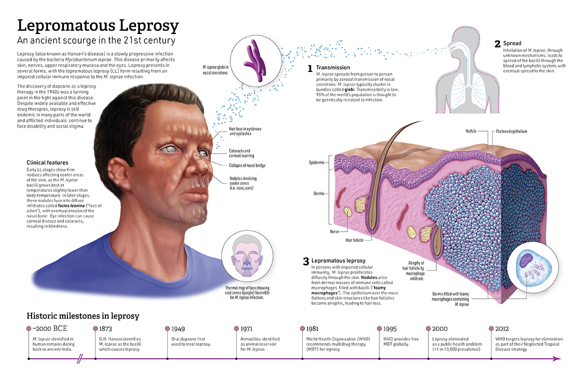
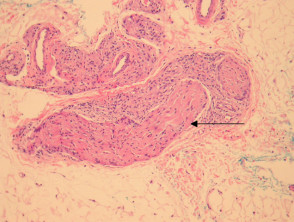
Bacteria are present in large numbers in cutaneous nerves and in endothelium and media of small and large vessels. Leprosy can lead to significant disability and stigmatisation. May have subcutaneous nodules erythema nodosum leprorum.
May invade arrectores pilorum muscle.

