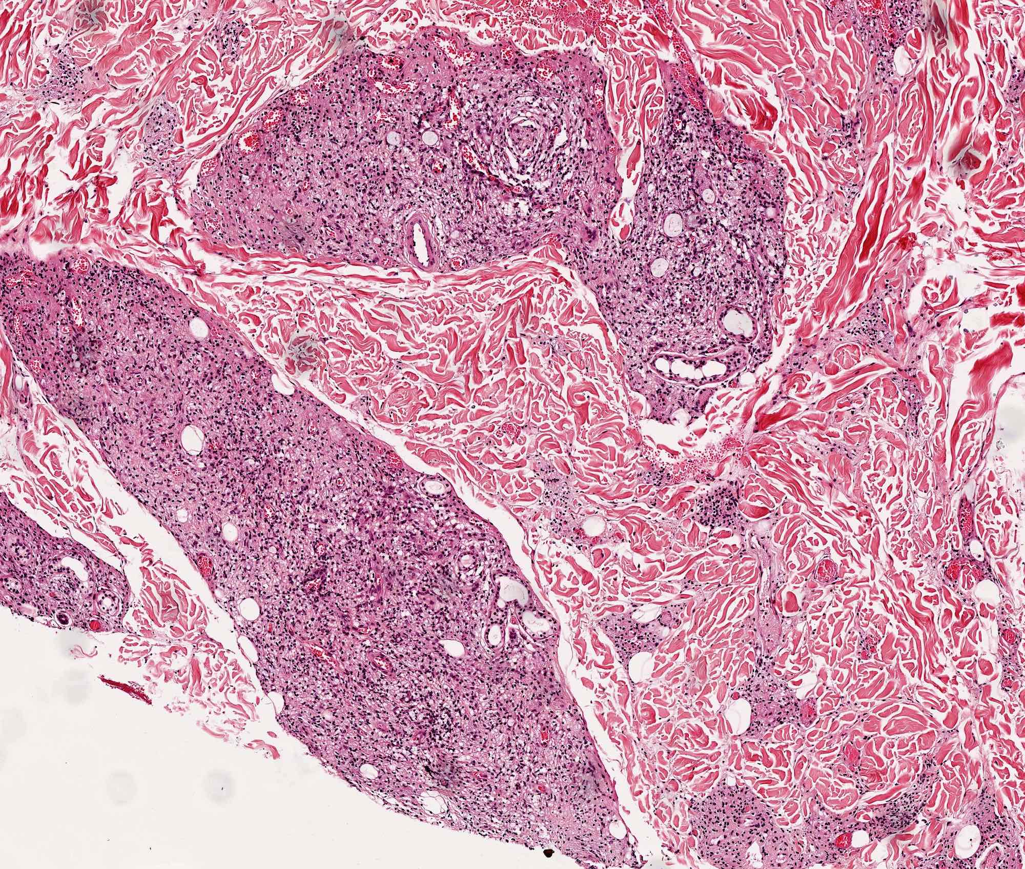
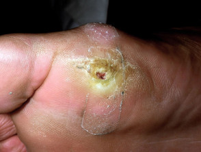
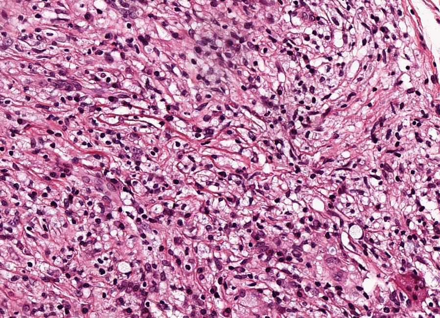
What Does Leprosy Look Like Under A Microscope
Leprae is well known in the lab scene for being a pain in the labcoat to work with.

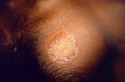
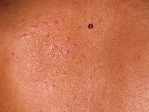
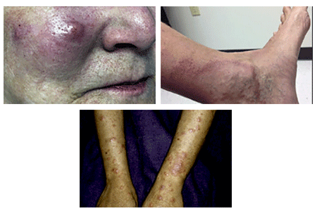
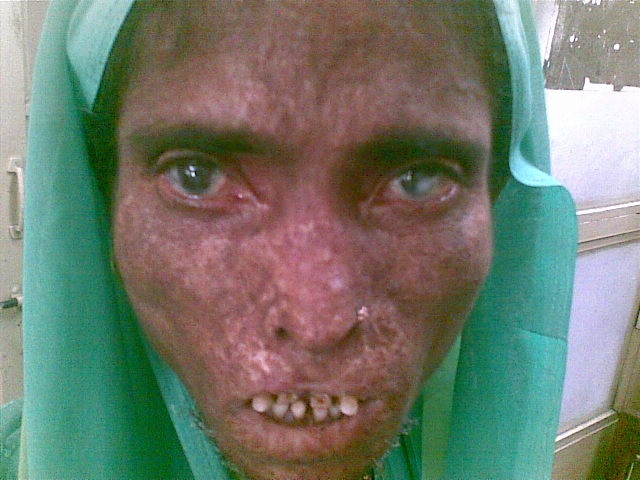
What does leprosy look like under a microscope. A small cut is made in the skin and a small amount of tissue fluid is taken. Leprosy symptoms this disease is characterized by the formation of skin bumps or lesions which looks like blister. Discover the symptoms and see pictures.
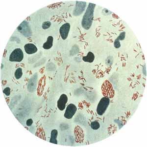
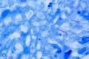
It is a strongly acid fast rod shaped organism with parallel sides and rounded ends. Another test used for diagnosis is a skin smear. Imaeda tamotsu instituto venezolano de investigaciones cientificas caracas venezuela and jacinto convitelectron microscope study of mycobacterium leprae and its environment in a vesicular leprous lesion.
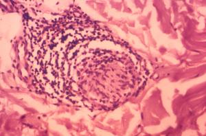
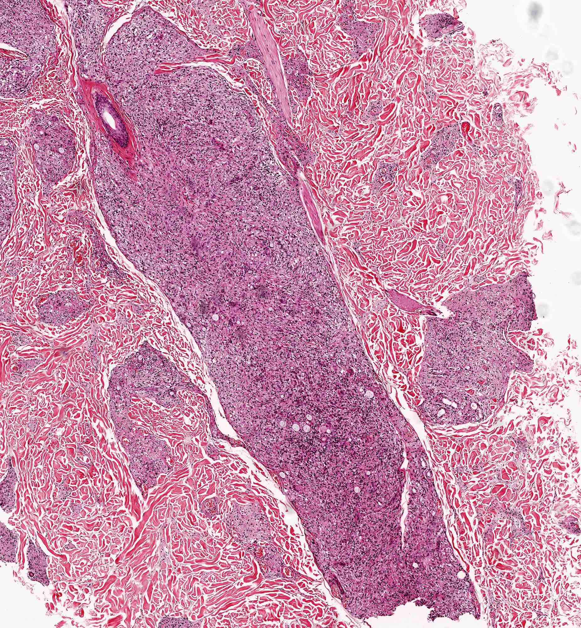
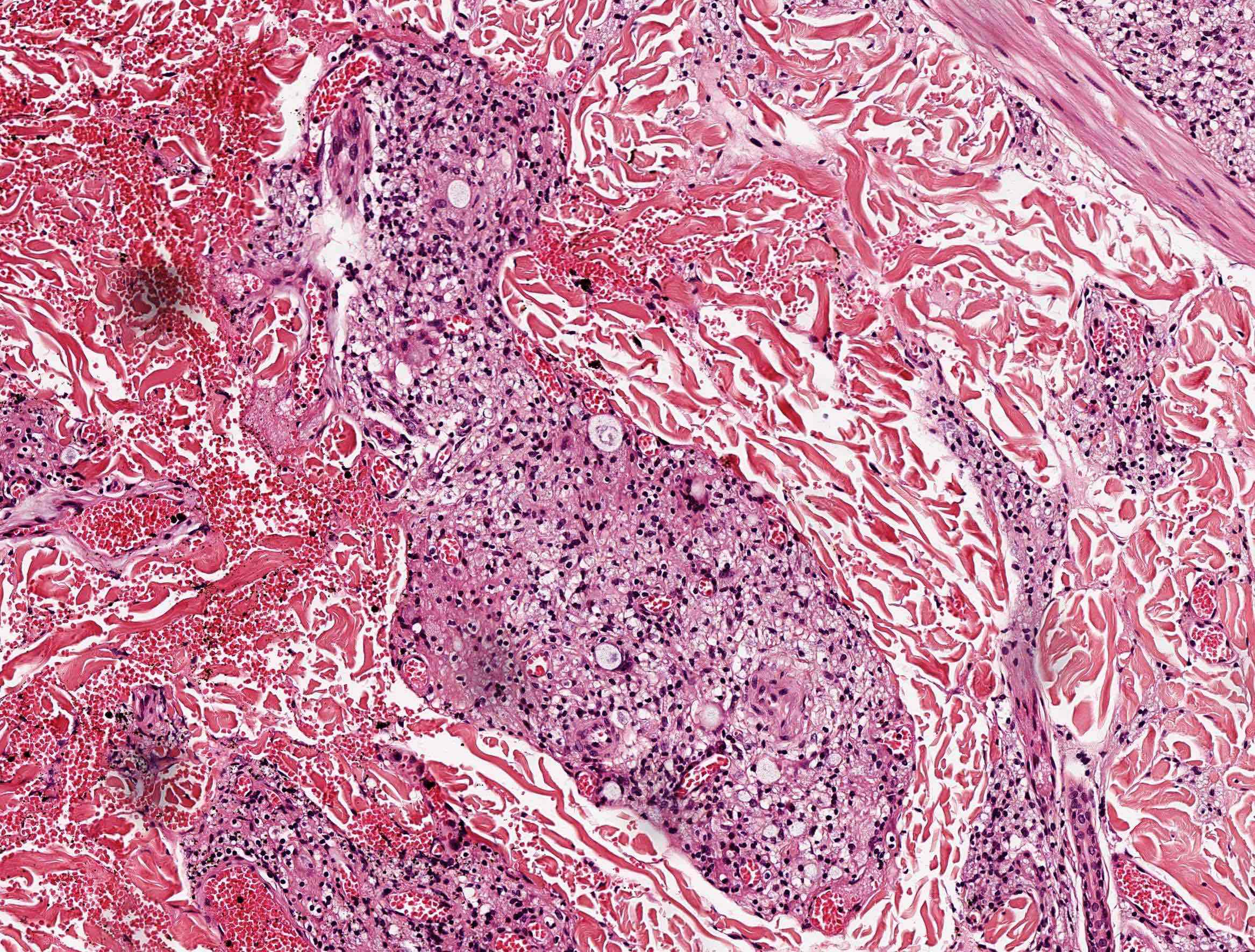
Tt leprosy is characterized by noncaseating granulomas destruction of dermal nerves loss of sweat glands and hair. Because leprosy can look like many other conditions your dermatologist may remove a bit of the afflicted skin or the fluid beneath it. Your dermatologist will also examine your skin.
The aetiological agent of leprosy is mycobacterium leprae. The image above was captured with a transmission electron microscope. Mycobacterium leprae is an acid fast rod shaped bacillus.
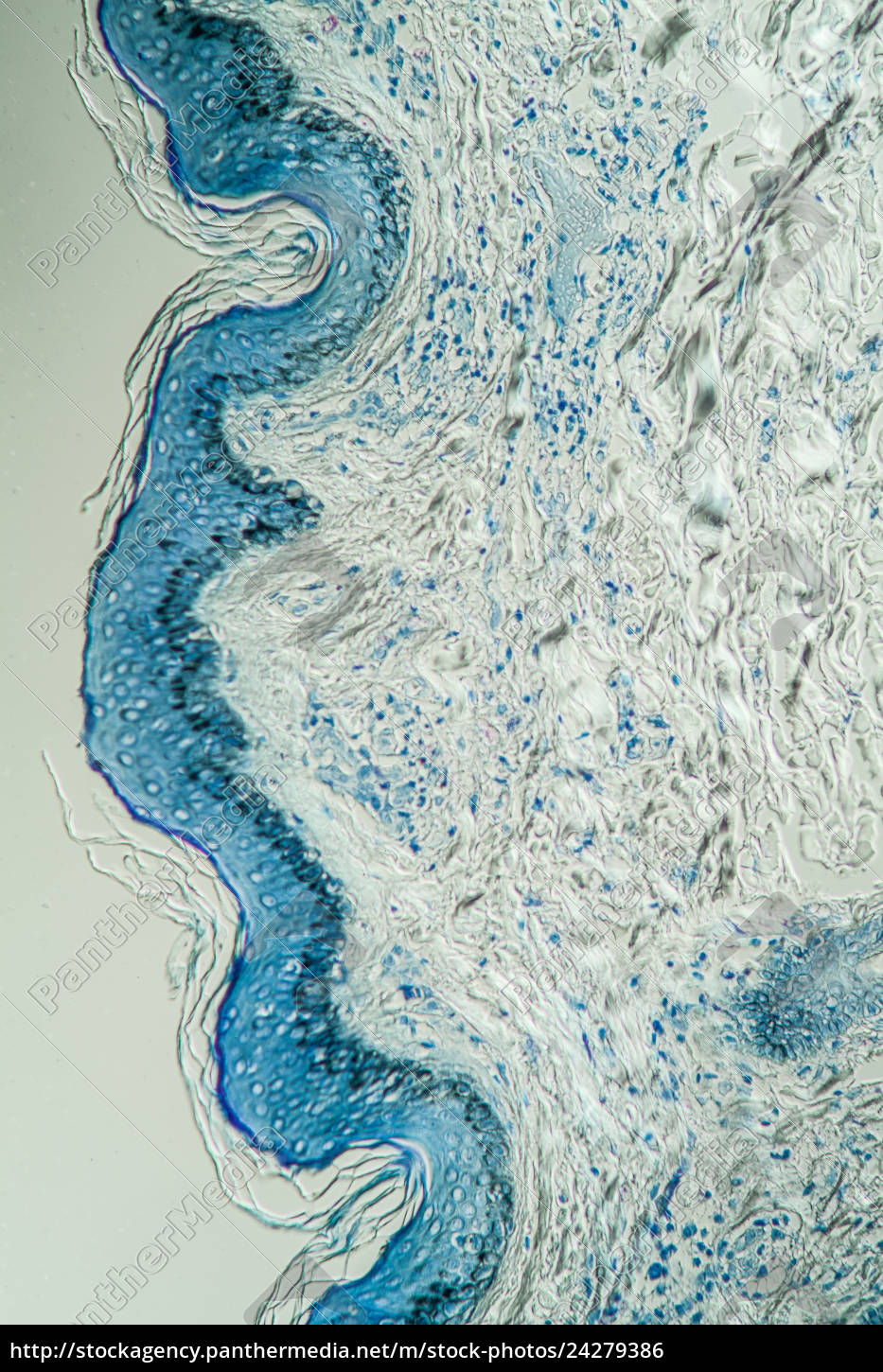
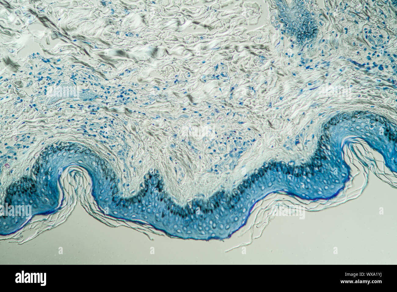
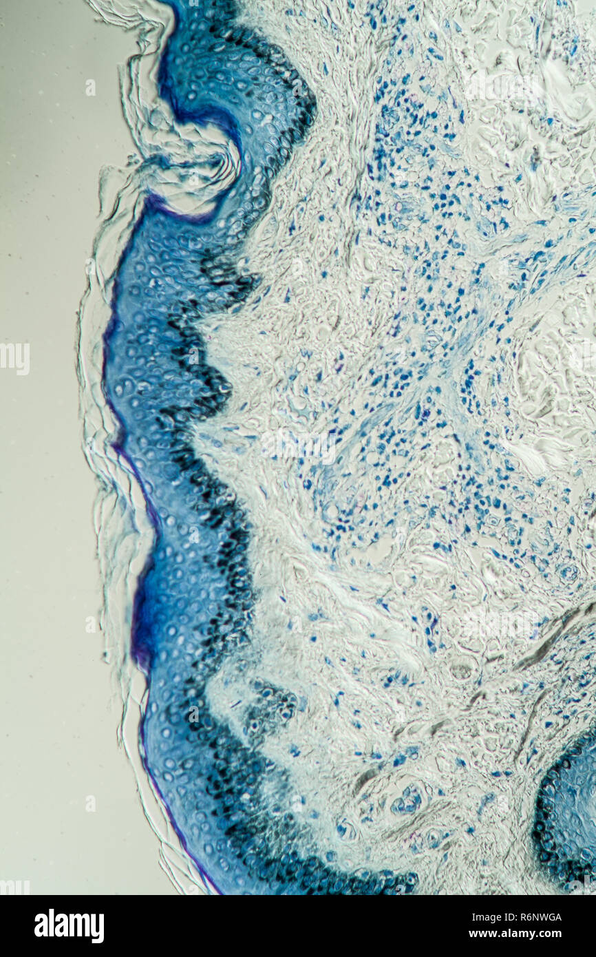
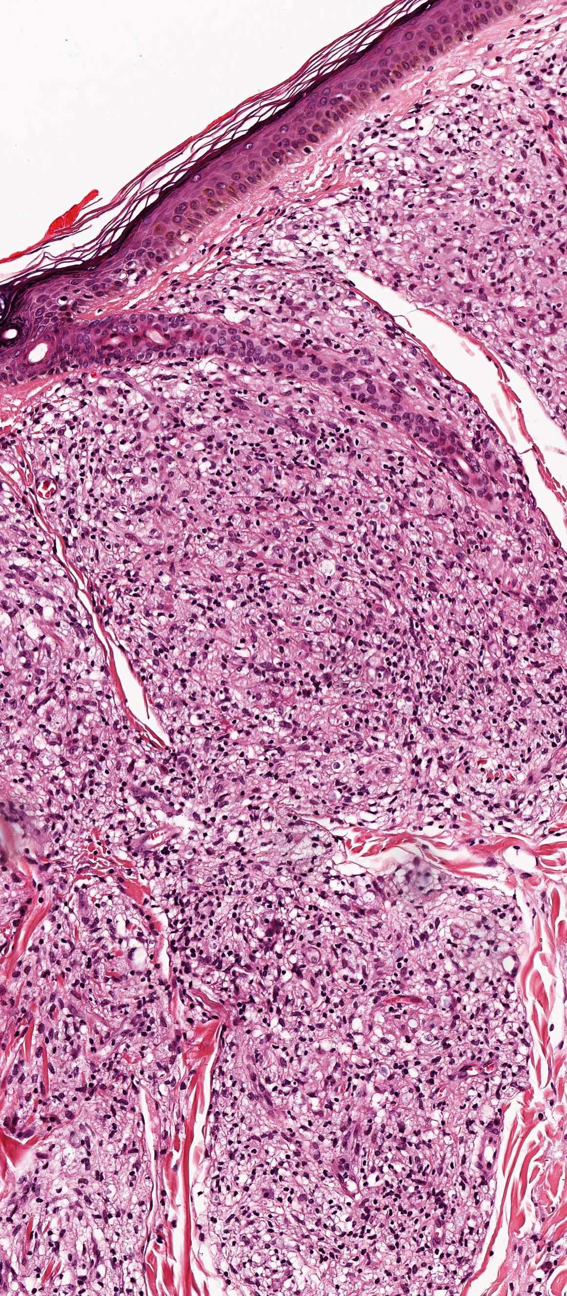
Histologic findings usually from skin biopsy vary based on the type of leprosy. 1962biopsied specimens of a borderline leprosy lesion were observed with the electron microscopein this lesion the majority of mycobacterium. Leprosy is a disease caused by a bacterial pathogen mycobacterium leprae.
It thrives at temperatures just under human body temperature and has a doubling time of almost 2 weeks where most other species double in a matter of hours. What is leprosy leprosy also known as hansens disease is a chronic infectious disease caused by slow growing bacteria called mycobacterium leprae which has a preference for the skin and peripheral nerves 1. This isnt quite as sharp as the first one but you can see the spikes on the surface of the virus which gives the coronavirus its name meaning crown.
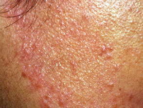
The skin color may change and the affected person may not be sensitive to heat or touch as the disease progresses. If the bacteria that cause leprosy are found the diagnosis is leprosy. This will be examined under a microscope.
Leprosy is diagnosed by taking a skin sample biopsy and examining it under the microscope looking for leprosy bacteria. Leprosy or hansens disease is a chronic progressive bacterial infection that can cause disfigurement and disability if left untreated. In size and shape it closely resembles the tubercle bacillus.
This is examined under a microscope for the presence of leprosy bacteria.






/cloudfront-ap-southeast-2.images.arcpublishing.com/nzme/UOECLLBW6MPNBEMTF6UZSEJ7MM.jpg)




/cloudfront-ap-southeast-2.images.arcpublishing.com/nzme/QPOE7EVGZTDWOMEJLSUEKLSDBU.jpg)



















/cdn.vox-cdn.com/uploads/chorus_image/image/64495794/1087411.0.jpg)


:max_bytes(150000):strip_icc()/GettyImages-181807923web-56f1f6d73df78ce5f83cbaaa.jpg)


