
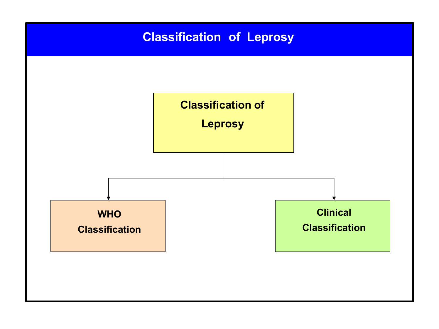
Paucibacillary And Multibacillary Leprosy Images

Source : commons.wikimedia.org
Source : www.scielo.org.co
Source : www.dinf.ne.jp

Source : www.researchgate.net

Source : www.cehjournal.org
Images 3 lab test 0 sidebars 1.
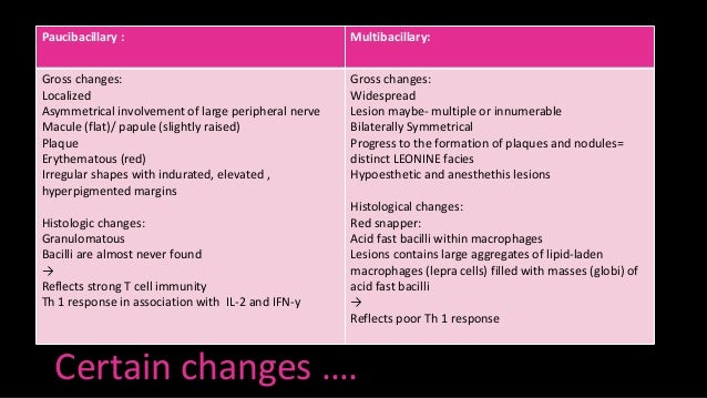
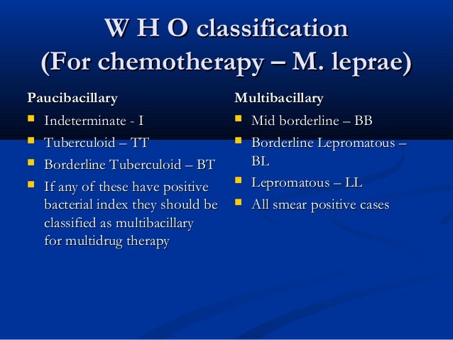
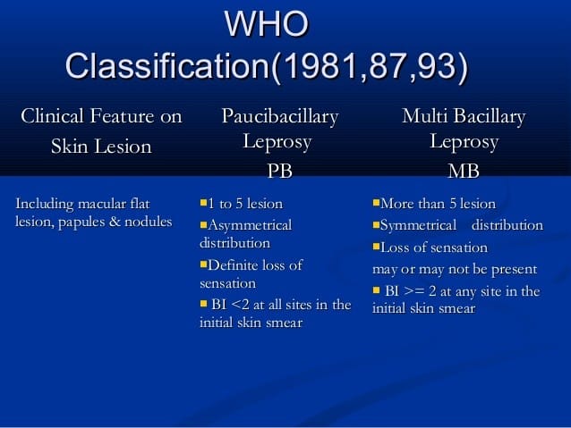
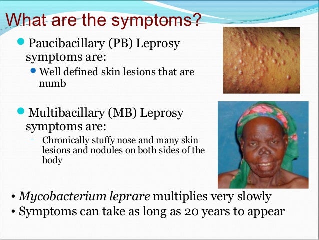
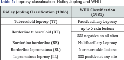
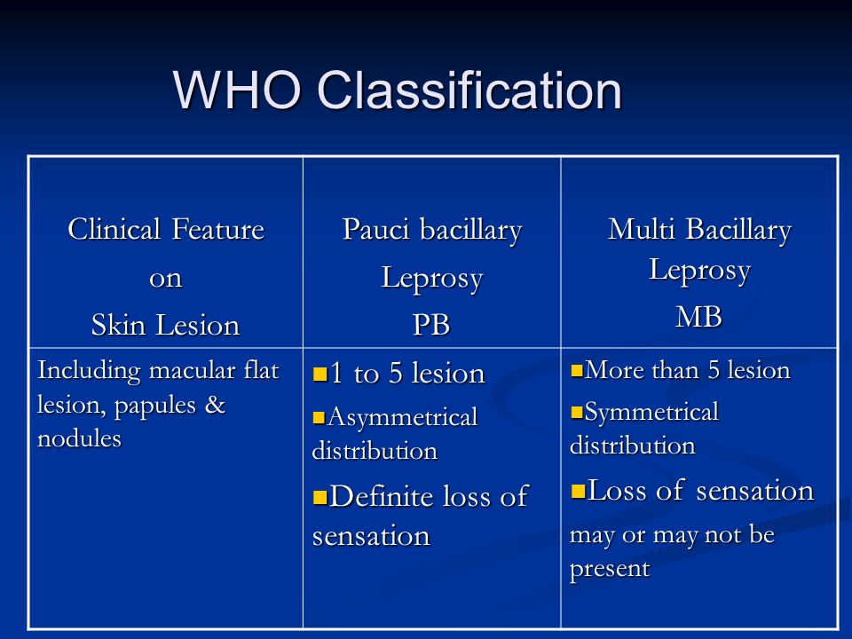
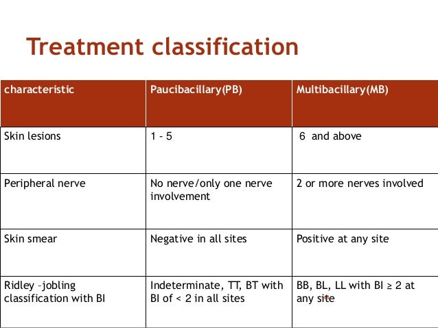
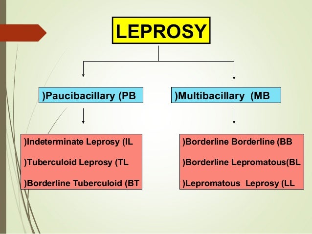
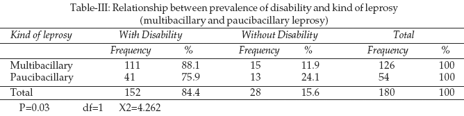
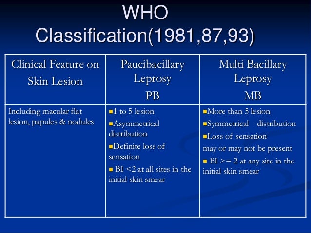
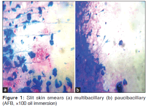
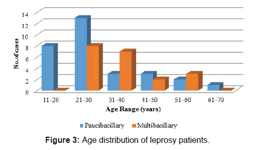
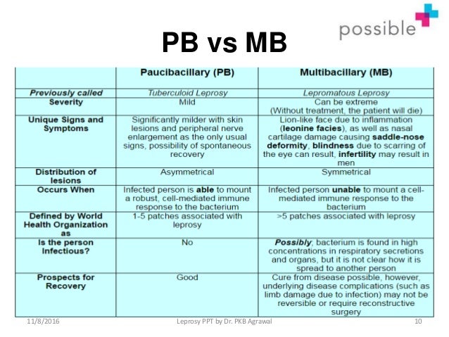
Paucibacillary and multibacillary leprosy images. In the classification based on skin smears patients showing negative smears at all sites are grouped as paucibacillary leprosy pb while those showing positive smears at any site are grouped as having multibacillary leprosy mb. Feb 6 2018 image result for paucibacillary leprosy vs multibacillary. Skin smears and biopsies.
People have multibacillary leprosy if they have 6 or more affected areas andor if bacteria are detected in a sample from an affected skin area. Explore searchviewparamsphrase by color family familycolorbuttontextcolorfamilyname related searches. In developing countries classification of multibacillary or paucibacillary leprosy is made by the combination of clinical examination findings and bacterial counts as determined on acid faststained smears made from skin slits of lesions and skin from cool areas of the body such as the earlobes.
Image result for paucibacillary leprosy vs multibacillary. No bacteria can be detected on samples from these areas. People with paucibacillary leprosy have 5 or fewer affected skin areas.
Feb 6 2018 image result for paucibacillary leprosy vs multibacillary. Leprosy can be classified on the basis of clinical manifestations and skin smear results. Ernst in goldmans cecil medicine twenty fourth edition 2012.
Source : studylib.net

Source : www.clinicalmicrobiologyandinfection.com
Source : www.x-mol.com
Source : journals.plos.org

Source : www.researchgate.net

Source : commons.wikimedia.org

Source : brainly.in
Source : www.webpathology.com
Source : www.actasdermo.org

Source : www.scielo.br

Source : www.ijpd.in
Source : pt.slideshare.net

Source : communitymedicine4all.com
Source : www.slideshare.net


Source : www.ijmyco.org
Source : www.worldofmedicalsaviours.com

Source : www.ijmyco.org

Source : slideplayer.com

Source : www.researchgate.net
Source : www.slideshare.net

Source : www.jaypeedigital.com

Source : www.cehjournal.org

Source : onlinelibrary.wiley.com
Source : juniperpublishers.com


Source : www.researchgate.net
Source : bmcinfectdis.biomedcentral.com

Source : www.researchgate.net

Source : www.scielo.org.mx

Source : www.sciencedirect.com

Source : pjms.com.pk
Source : slideplayer.com
Source : www.intechopen.com

Source : internationaltextbookofleprosy.org

Source : www.youtube.com

Source : publichealth.choate.edu

Source : www.researchgate.net

Source : www.jaypeedigital.com
Source : www.alamy.com

Source : www.pinterest.com

Source : larvanator.weebly.com
Source : www.slideshare.net
Source : pjms.com.pk
Source : juniperpublishers.com

Source : www.e-ijd.org

Source : www.jpma.org.pk


Source : www.semanticscholar.org

Source : encrypted-tbn0.gstatic.com

Source : communitymedicine4all.com

Source : www.ijpd.in

Source : www.pharmacy180.com

Source : www.semanticscholar.org
Source : www.intechopen.com

Source : www.learnscience.info
Source : oldfiles.bjorl.org

Source : www.semanticscholar.org

Source : web.stanford.edu
Source : encrypted-tbn0.gstatic.com
Source : www.open.edu
Source : encrypted-tbn0.gstatic.com

Source : www.semanticscholar.org
Source : www.slideshare.net
Source : juniperpublishers.com
Source : www.slideshare.net
Source : twitter.com
Source : www.slideshare.net
Source : link.springer.com
Source : encrypted-tbn0.gstatic.com
Source : www.amhsr.org
Source : www.slideshare.net
Source : journals.plos.org
Source : www.alamy.com
Source : www.amhsr.org
Source : www.amhsr.org
Source : www.slideshare.net

Source : journals.plos.org

Source : www.semanticscholar.org

Source : web.stanford.edu

Source : www.dinf.ne.jp

Source : en.wikipedia.org

Source : www.researchgate.net
Source : communitymedicine4all.com