Lepra Cells Seen In
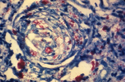
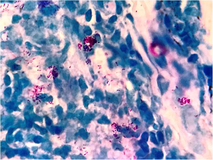
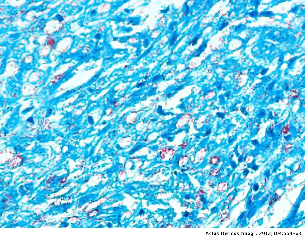
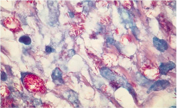
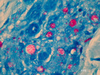
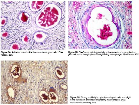
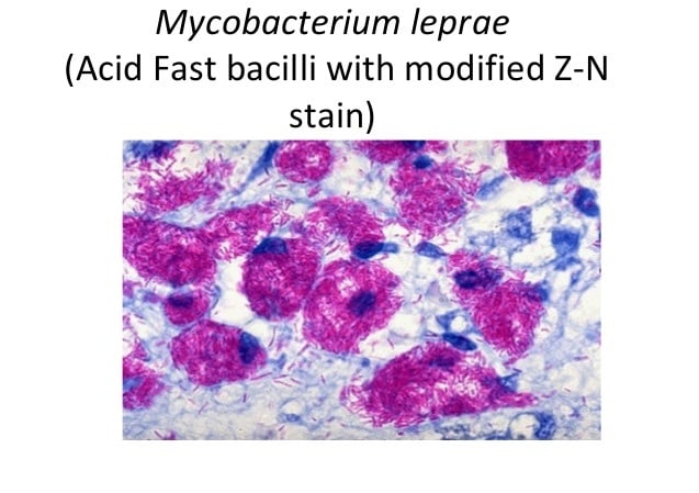
It occurs in large numbers in the lesions of lepromatous leprosy chiefly in masses within the lepra cells often grouped together like bundles of cigars or arranged in a palisade.
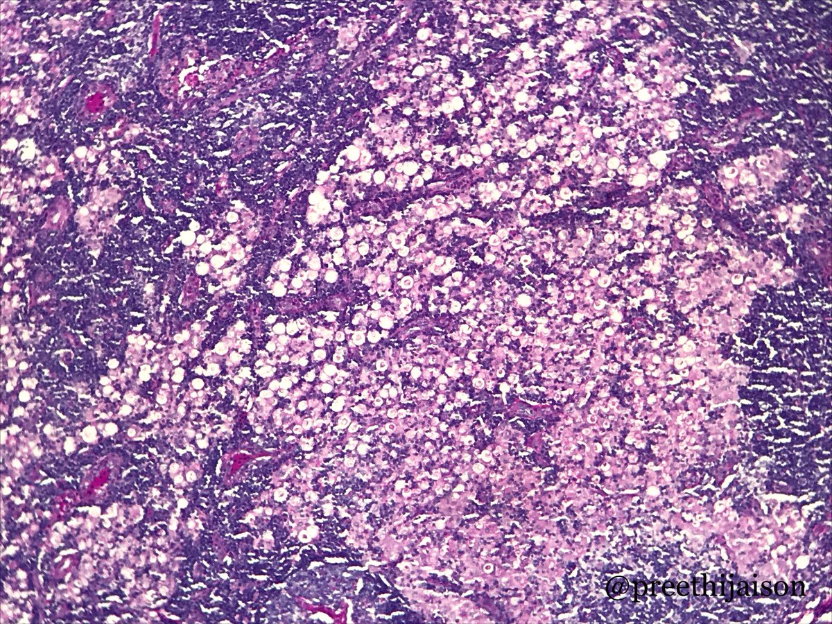
Lepra cells seen in. Known as multibacillary disease. Here you will find more than 5000. Abnormalities in endothelial cell structure occur with resulting breaches in the blood nerve barrier.
Chains are never seen. Foam cells are macrophages that have ingested or phagocytized m. Inflammation immunity and hypersensitivity inflammation immunity and hypersensitivity medical mcqs medical mcqs for exams preparation of medical students and professionals.
Not readily contagious it. The infection is currently found in over 100 countries often located in high burden areas against a low burden background of cases. Leprae bacteria but are unable to digest the organisms who in turn multiply and use the macrophage as a method of transport throughout the body.
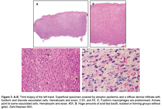
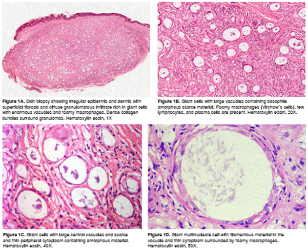
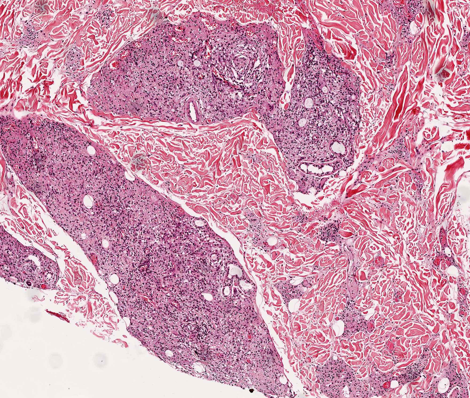
Most striking are the intracellular and extra cellular masses known as globi which consist of clumps of bacilli in capsular material. Bacilli seen in large numbers as globi inside lepra cells or extracellularly. As part of the human immune response white blood cell derived macrophages may engulf m.
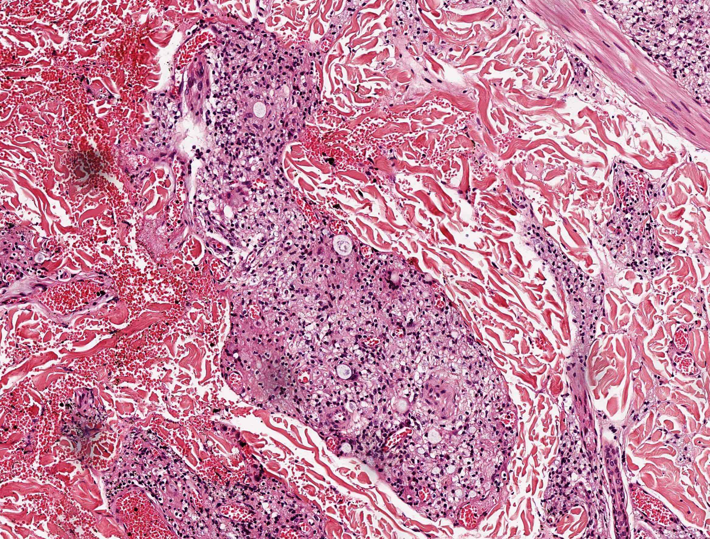
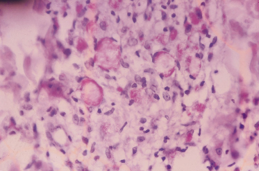
May have subcutaneous nodules erythema nodosum leprorum. Macrophages may be distended with large groups of leprosy bacilli globi. May invade arrectores pilorum muscle.
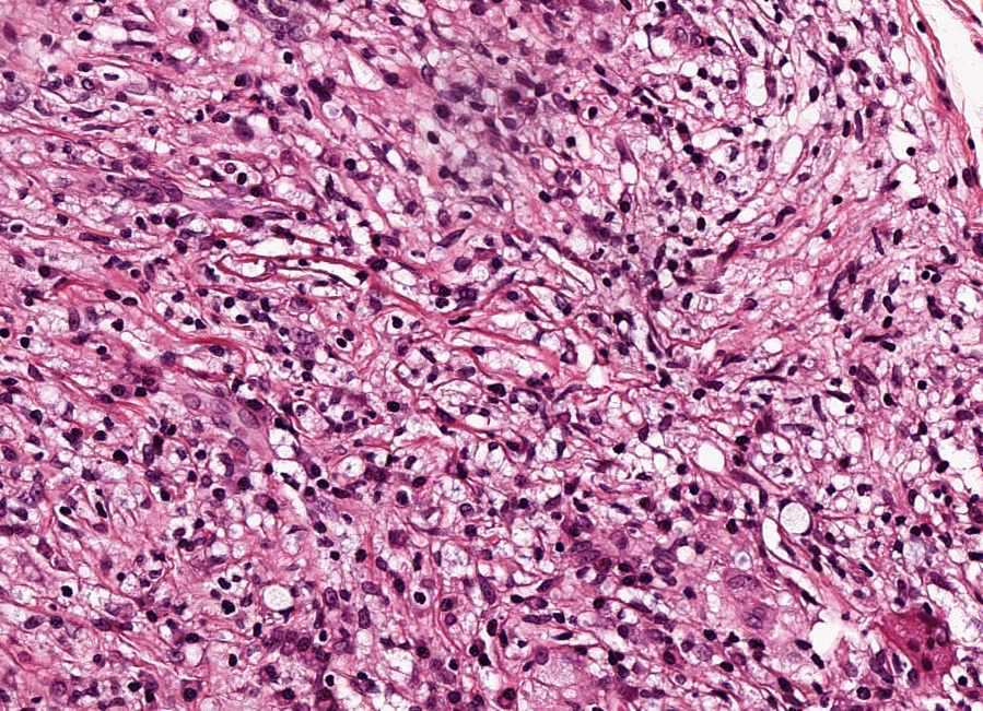
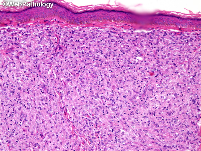
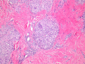
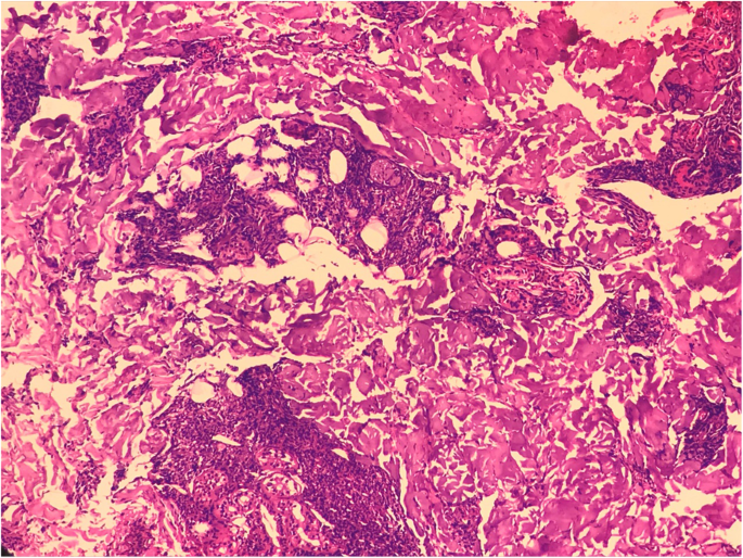
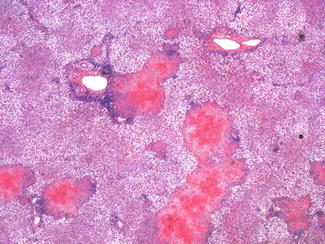
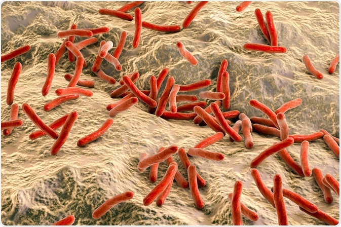
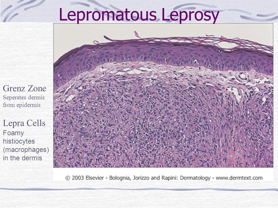
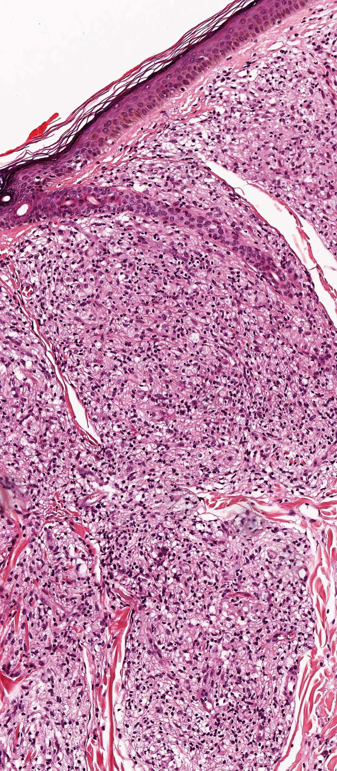
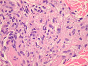
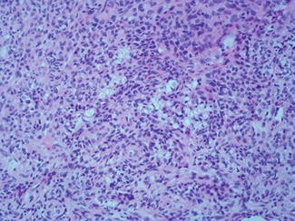
Bacteria are present in large numbers in cutaneous nerves and in endothelium and media of small and large vessels. Macrophages virchow cells lepra are found in poorly circumscribed masses in the dermis with few no lymphocytes. Mycobacterium leprae is the aetiologic agent of leprosy affecting the skin and peripheral nerves.
Lepra cells seen in leprosy are. 7 even in long standing cases of lepromatous disease the fascicular structure of nerve trunks remains discernible. Leprosy lepro se an inflammatory disease caused by mycobacterium leprae manifested in various ways depending on the hosts ability to develop cell mediated immunity.
As the disease progresses lesions infiltrate forming plaques and. Many can be seen in endothelium of endoneurial blood vessels and occasionally in cells within the lumen of such vessels. In the initial stages small sensory and autonomic nerve fibers in the skin of a person with leprosy are damaged.
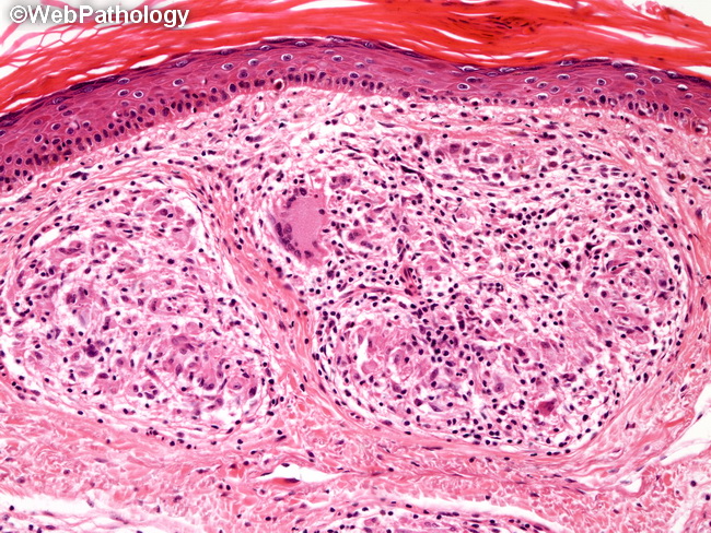
It is a chronic communicable disease characterized by the production of granulomatous lesions of the skin mucous membranes and peripheral nervous system. Distinctive large mononuclear phagocytes macrophages with a foamlike cytoplasm and also poorly staining saclike structures resulting from degeneration of such cells observed characteristically in leprous inflammatory reactions. Lepromatous seen where host resistance is low.
Indistinct staining results from numerous fairly closely packed leprosy bacilli which are acid fast and resistant to staining by ordinary methods but may be vividly demonstrated by acid fast staining procedures. Gillis in molecular medical microbiology second edition 2015. Antibodies are produced but they do not prevent bacterial proliferation.


