
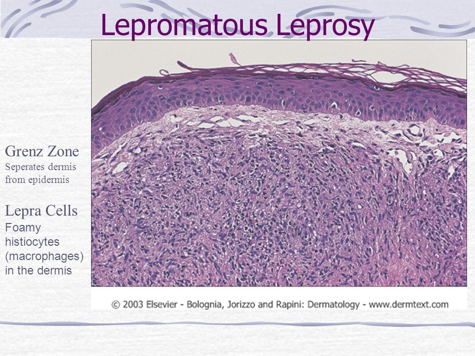
Lepra Cells Histology

Source : internationaltextbookofleprosy.org


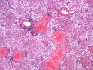
Source : www.scielo.br
Source : philippinejournalofpathology.org

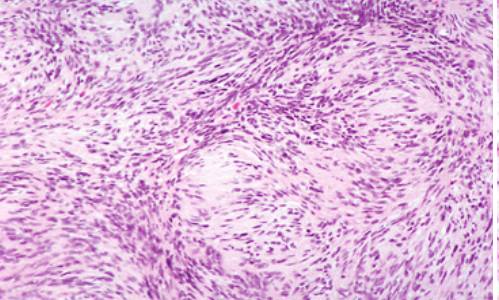
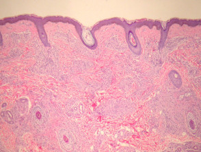
Occur in patients with borderline forms of leprosy.

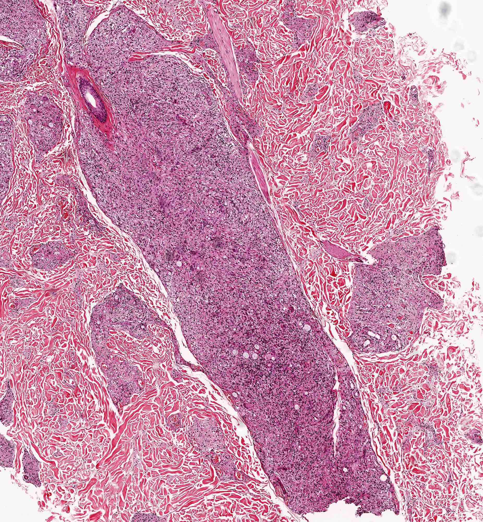
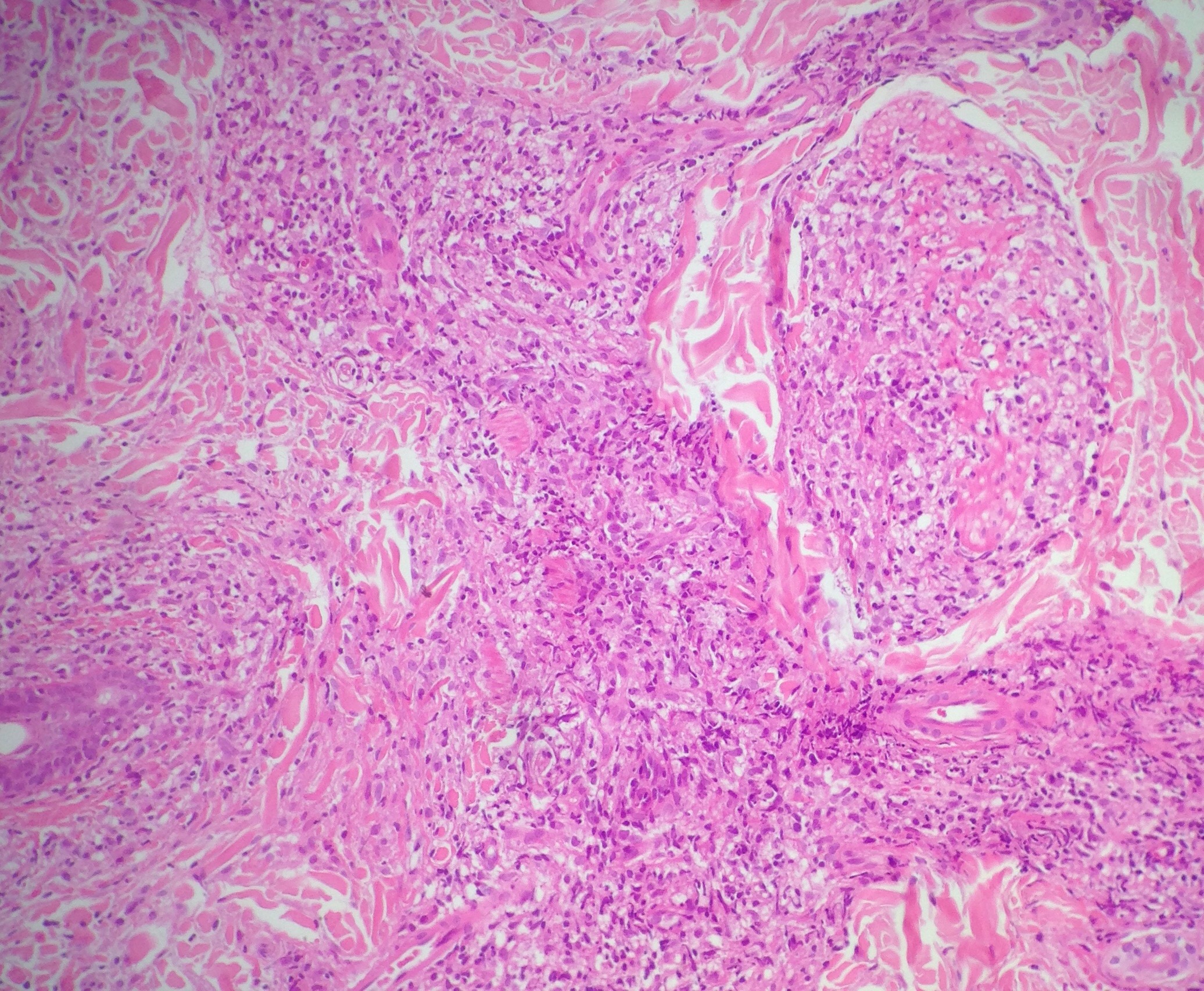
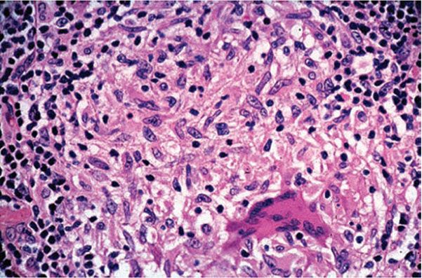
Lepra cells histology. The aetiological agent of leprosy is mycobacterium leprae. The majority of patients however fall into a broad dimorphous or borderline category between the two polar forms. The associated infiltrate is usually sparse and mainly lymphocytic.
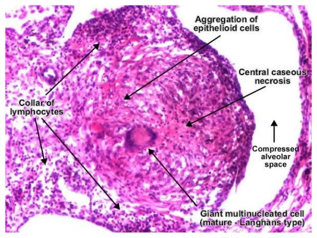
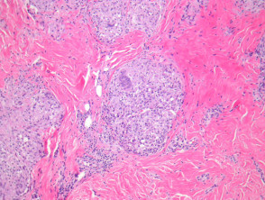
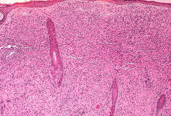
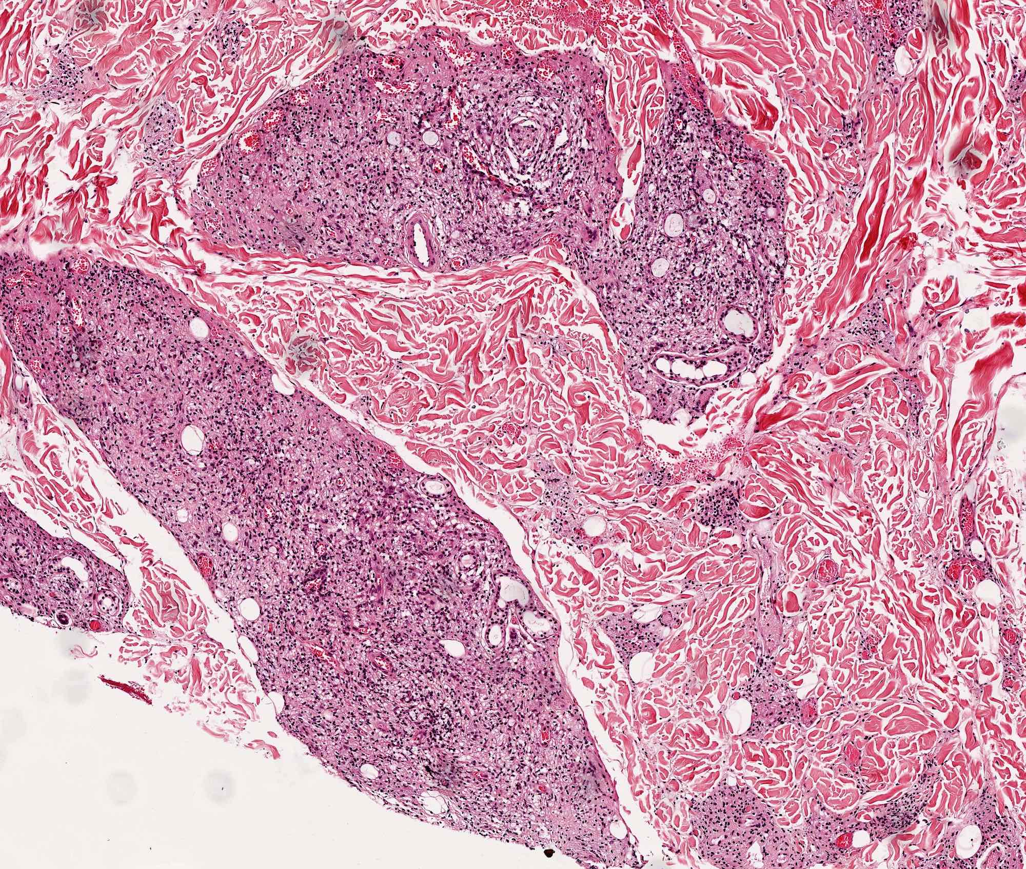
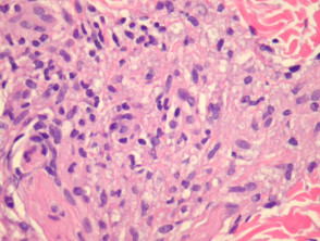
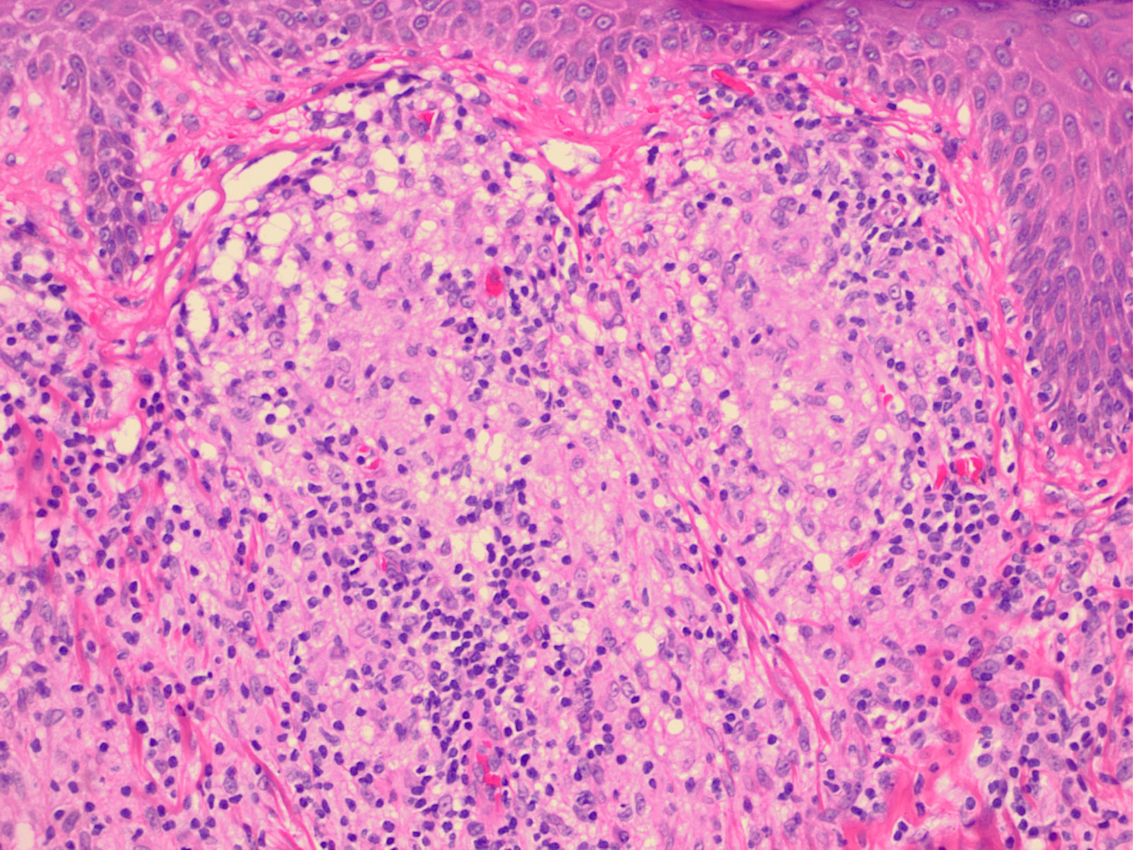
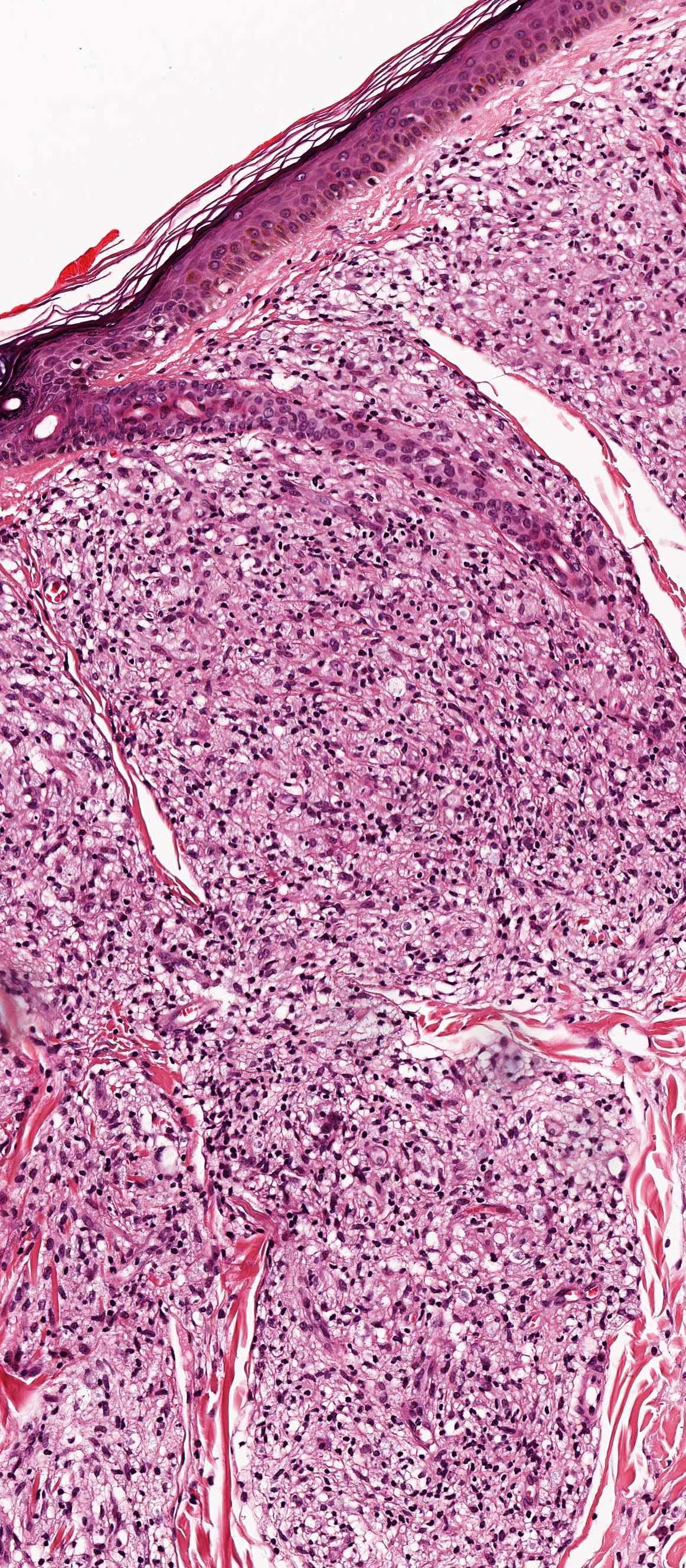
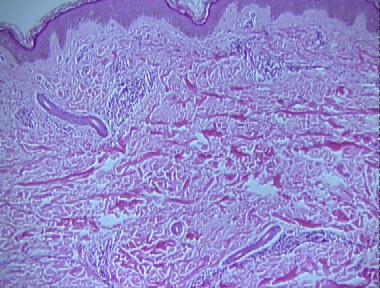
Histopathology of type 1 lepra reaction shows edema within granuloma separating inflammatory cells with density of lymphocytes in the dermis. Mainly involves the trunk. It examines the correlation between structure and function.
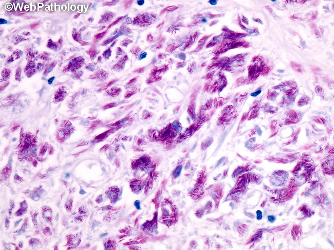
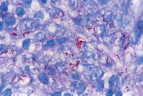
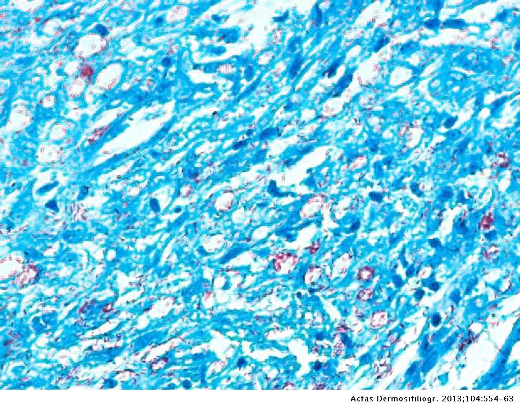
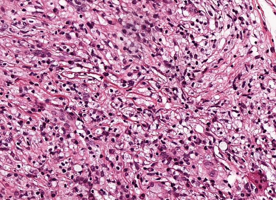
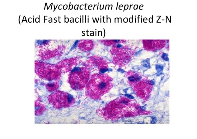
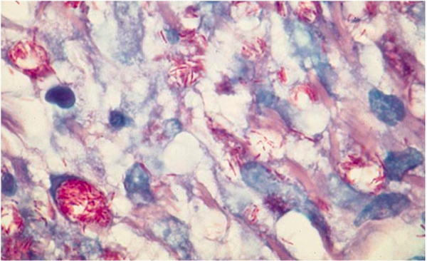
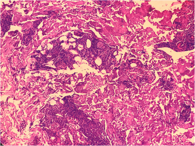
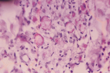
Type 1 lepra reactions. It occurs in large numbers in the lesions of lepromatous leprosy chiefly in masses within the lepra cells often grouped together like bundles of cigars or arranged in a palisade. In size and shape it closely resembles the tubercle bacillus.
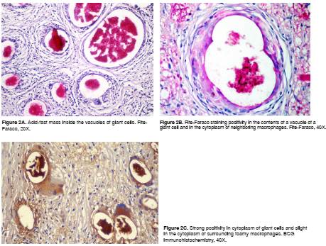
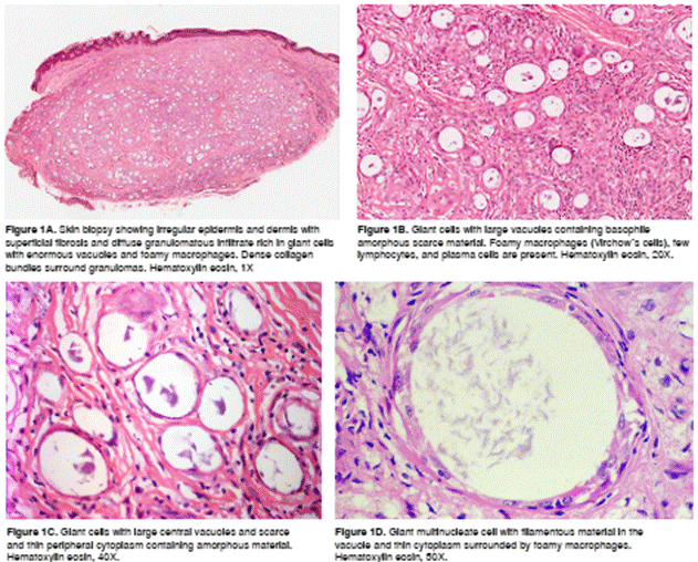
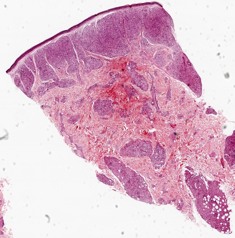
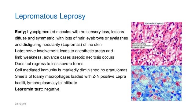
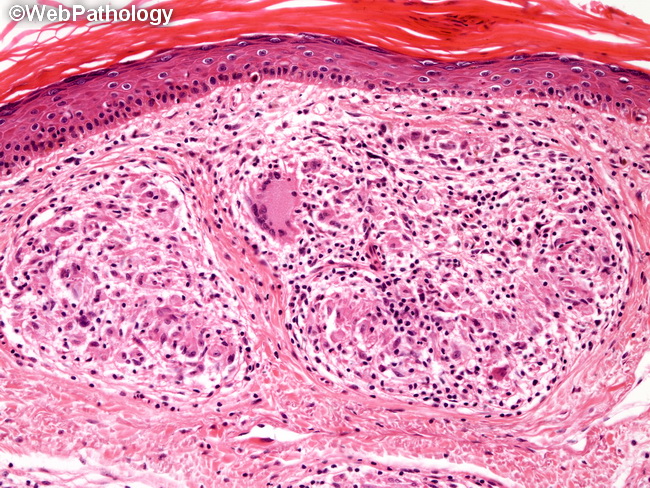
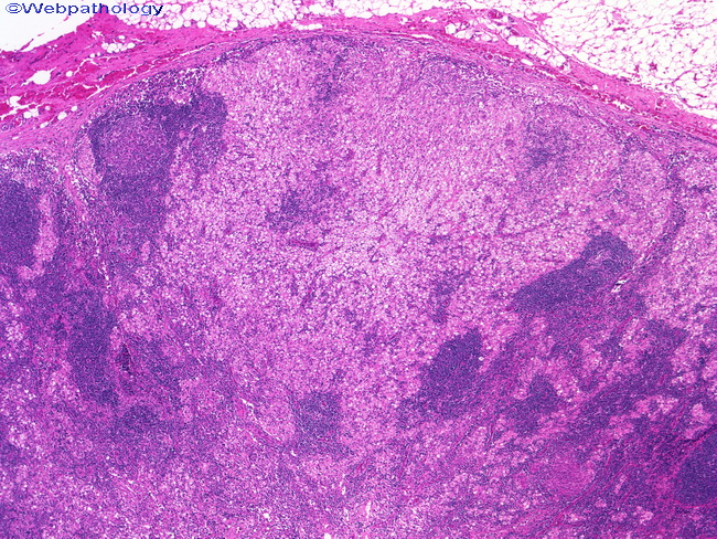
Lepromatous leprosy underneath a normal epidermis and grenz zone there are sheets or clusters of macrophages figure 1. The abundance of foamy cells may be mistaken for some form of histiocytosis if leprosy is not suspected and if fite stains are not done to demonstrate the numerous acid fast organisms. Occasionallythere is appearance of new skin lesionsneuritis and fever low grade.
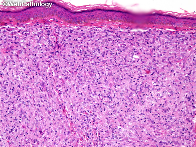
Classic signs of inflammation within previously involved maculespapules and plaqueswhich are markedly erythematous swollen and oedematous. Histology is the study of the microanatomy of cells tissues and organs as seen through a microscope. Pathology pathology mcqs for preparation.
Leprae resistant highly infected form of leprosy is termed polar lepromatous ll. It is delayed type of hypersensitivity. Histology guide teaches the visual art of recognizing the structure of cells and tissues and understanding how this is determined by their function.
Pathology multiple choice questions mcq for entrance examinations and other competitive examinations for all experienced freshers and students. Associated with large numbers of t cells bearing receptors a unique feature of leprosy. It is a strongly acid fast rod shaped organism with parallel sides and rounded ends.
Langerhans cell histiocytosis usually occurs in young children cells have pale pink cytoplasm and bean shaped nuclei eosinophils are usually prominent acid fast stain and microbiologic studies negative cells are s 100 protein and cd1a positive pneumocystis carinii extracellular foamy exudate gms or immunohistochemical stain highlight the organisms.
Source : philippinejournalofpathology.org
Source : encrypted-tbn0.gstatic.com
Source : encrypted-tbn0.gstatic.com
Source : www.pathologyoutlines.com
Source : www.webpathology.com

Source : www.epathologies.com
Source : www.slideshare.net
Source : dermnetnz.org
Source : www.scielo.org.co

Source : www.dermatologyadvisor.com

Source : www.dermaamin.com

Source : www.semanticscholar.org

Source : www.hindawi.com

Source : www.ijmyco.org

Source : www.researchgate.net
Source : www.pathologyoutlines.com

Source : histopath.wordpress.com

Source : www.actasdermo.org
Source : encrypted-tbn0.gstatic.com

Source : onlinelibrary.wiley.com
Source : www.actasdermo.org

Source : www.hindawi.com

Source : www.epathologies.com
Source : www.pathologyoutlines.com
Source : www.memorangapp.com
Source : www.scielo.org.co
Source : slideplayer.com
Source : www.pathologyoutlines.com
Source : www.pathologyoutlines.com

Source : www.histopathology-india.net


Source : internationaltextbookofleprosy.org

Source : medicoapps.org

Source : internationaltextbookofleprosy.org
Source : microbenotes.com

Source : www.ajmhs.org

Source : www.histopathology-india.net
Source : revistabiomedica.org

Source : www.ijdvl.com
Source : dermnetnz.org
Source : histopath.wordpress.com

Source : www.histopathology-india.net

Source : www.epathologies.com
Source : www.slideshare.net

Source : www.researchgate.net

Source : www.sciencedirect.com

Source : www.ijdvl.com

Source : www.jaypeedigital.com

Source : www.epathologies.com

Source : idoj.in

Source : www.frontiersin.org

Source : onlinelibrary.wiley.com

Source : imedscholar.com

Source : www.dermatologyadvisor.com
Source : www.pathologyoutlines.com

Source : internationaltextbookofleprosy.org

Source : medicoapps.org

Source : www.researchgate.net


Source : www.ctdt.co.in
Source : encrypted-tbn0.gstatic.com

Source : www.jaypeedigital.com
Source : plasticsurgerykey.com
Source : dermaamin.com

Source : www.mendeley.com

Source : www.assaygenie.com
Source : www.webpathology.com

Source : www.sciencedirect.com

Source : www.researchgate.net

Source : histopath.wordpress.com
Source : www.memorangapp.com
Source : dermnetnz.org
Source : www.webpathology.com


Source : www.facebook.com
Source : www.histopathology-india.net

Source : jmedicalcasereports.biomedcentral.com
Source : jmedicalcasereports.biomedcentral.com
Source : www.pathologyoutlines.com

Source : www.e-ijd.org

Source : www.redalyc.org
Source : www.dxpath.com

Source : www.scielo.br
Source : plasticsurgerykey.com
Source : www.webpathology.com

Source : www.e-ijd.org

Source : www.researchgate.net


Source : internationaltextbookofleprosy.org