Lepra Cells Found In Lepromatous Leprosy Are
Of cd8 t cells.

Lepra cells found in lepromatous leprosy are. May invade arrectores pilorum muscle. Plasma cells are found. Sadaf fasih dermatology deptt a man with deformities of hands loss of sensations and chronic skin lesions leprosy chronic granulomatous disease caused by mycobacterium lepraeprincipally affecting peripheral nerves skin prevalence 600000 new casesyear worldwide 23rd of worlds leprosy burden in asia espin indiapakistannepalbangladesh tanzania.
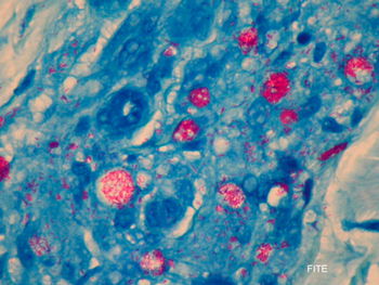
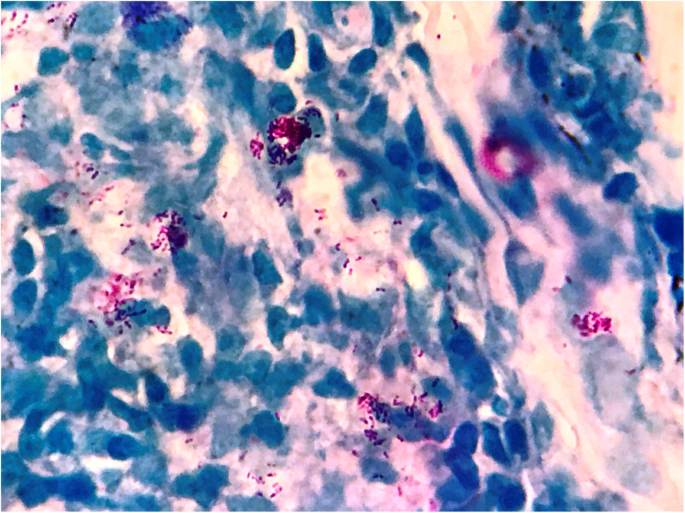
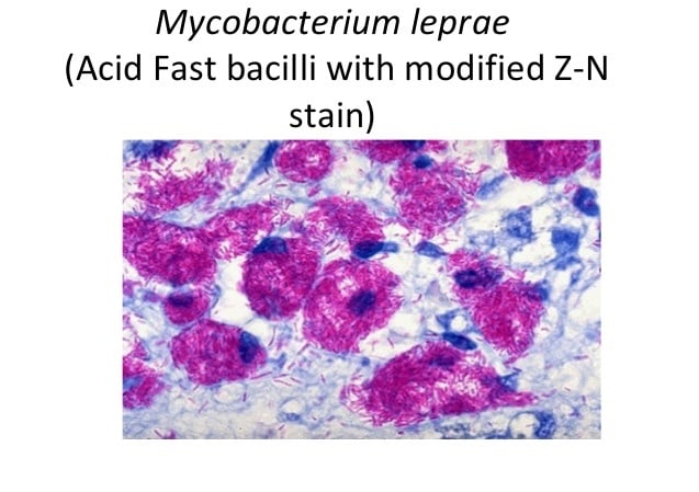
Indistinct staining results from numerous fairly closely packed leprosy bacilli which are acid fast and resistant to staining by ordinary methods. This is the most unfavorable clinical variant of leprosy. Lepromatosis is a relatively newly identified mycobacterium isolated from a fatal case of diffuse lepromatous leprosy in 2008.
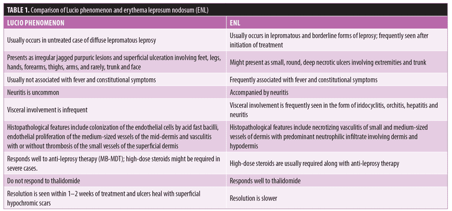
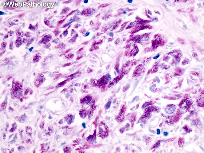
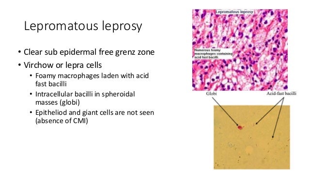
May have subcutaneous nodules erythema nodosum leprorum. And many intracellular afb which are frequently found in globi. Dermal nerves are easily visible.
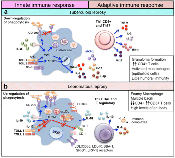
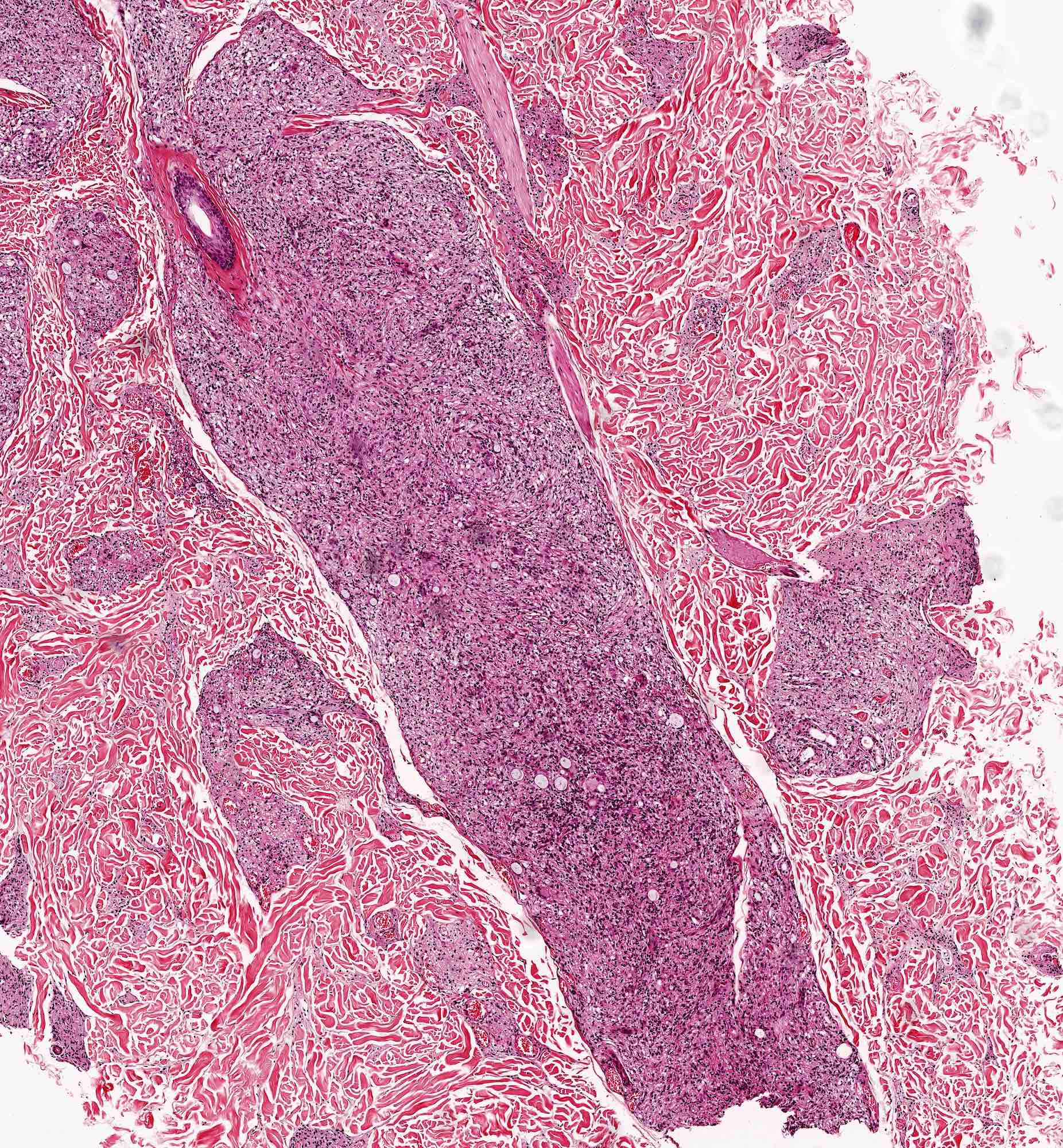
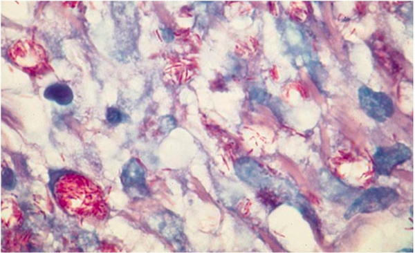
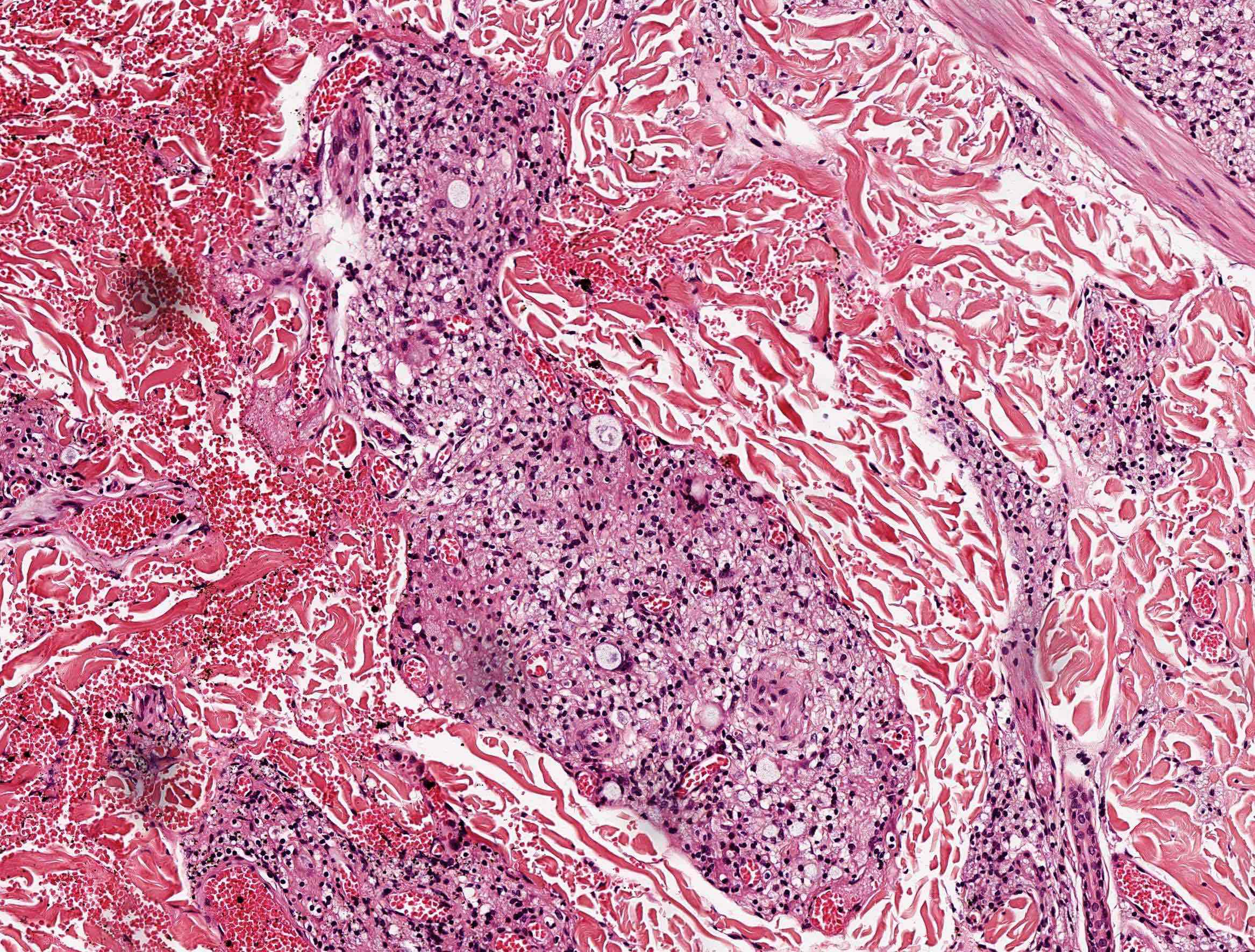
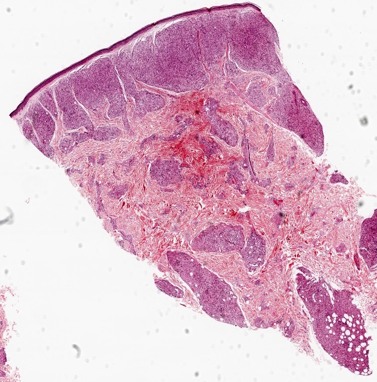
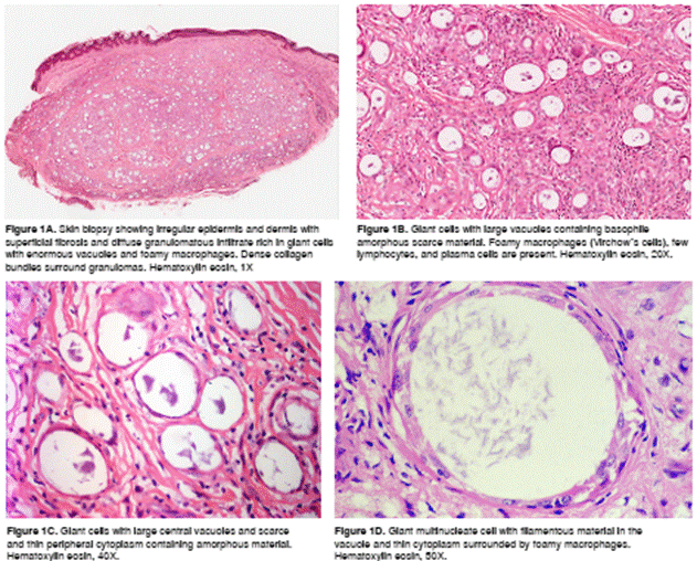
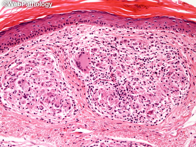
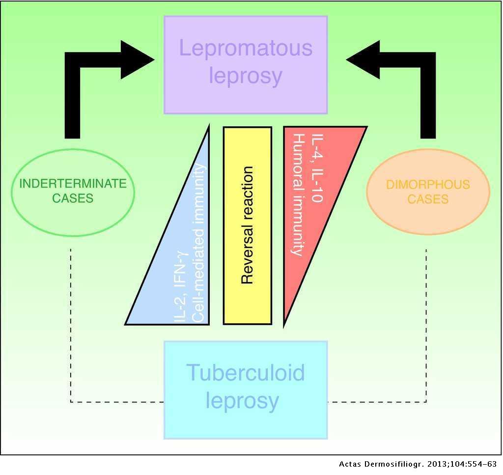
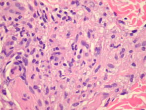
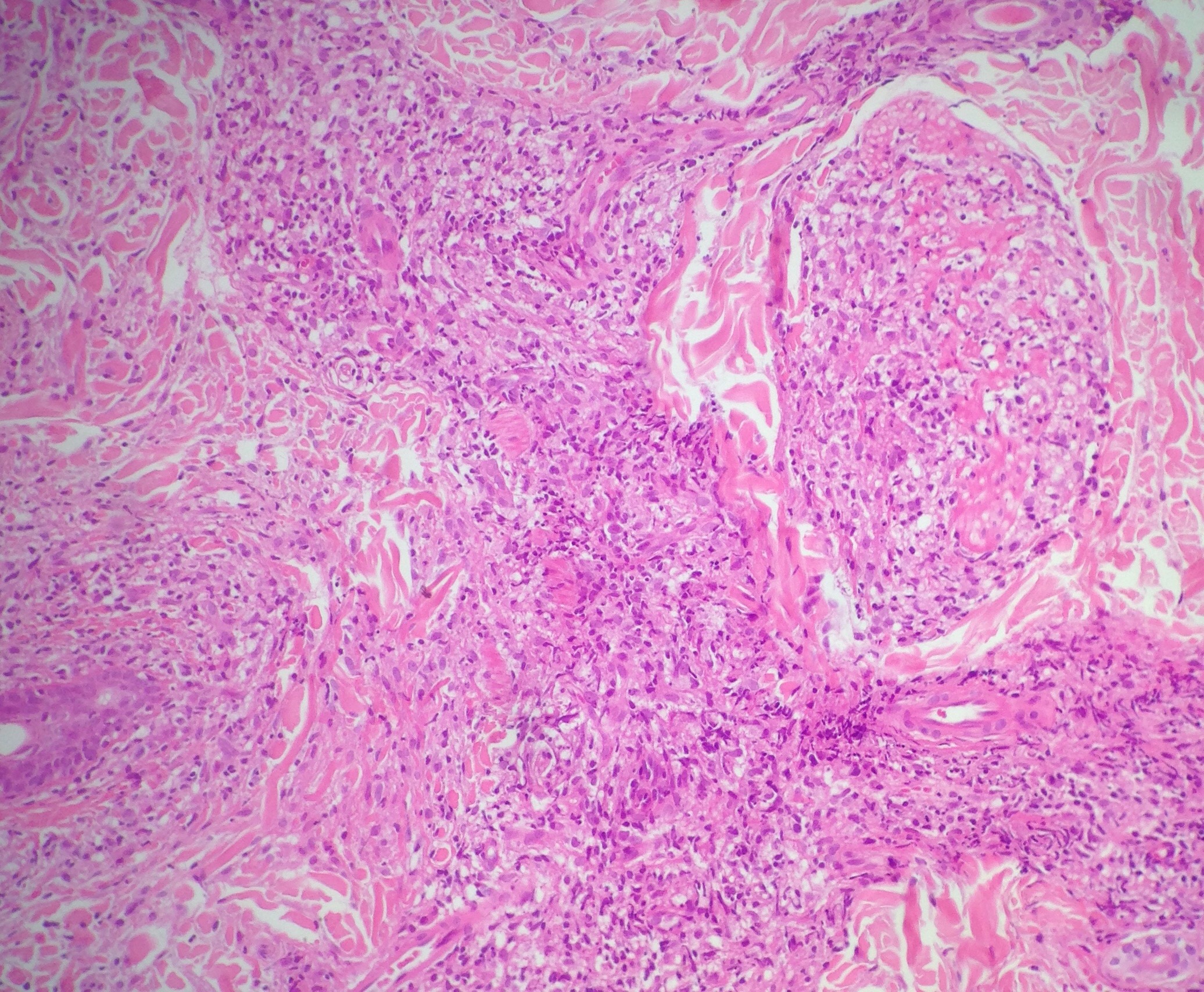
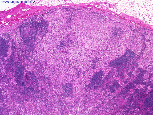
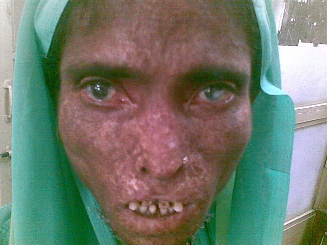
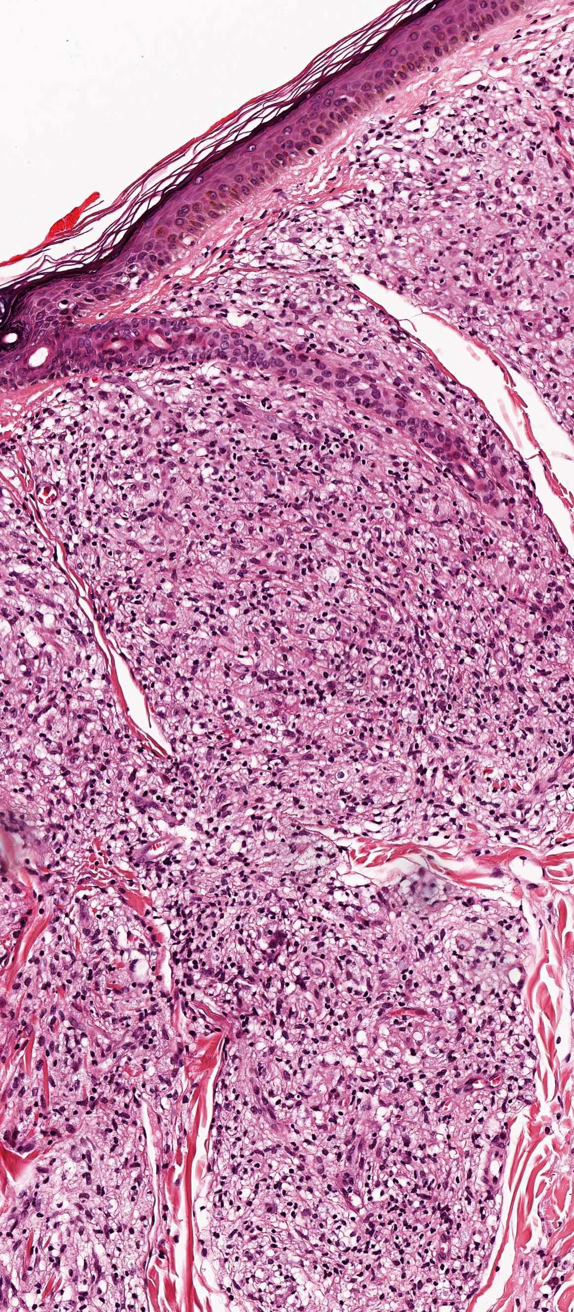
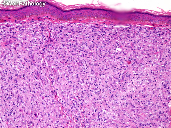
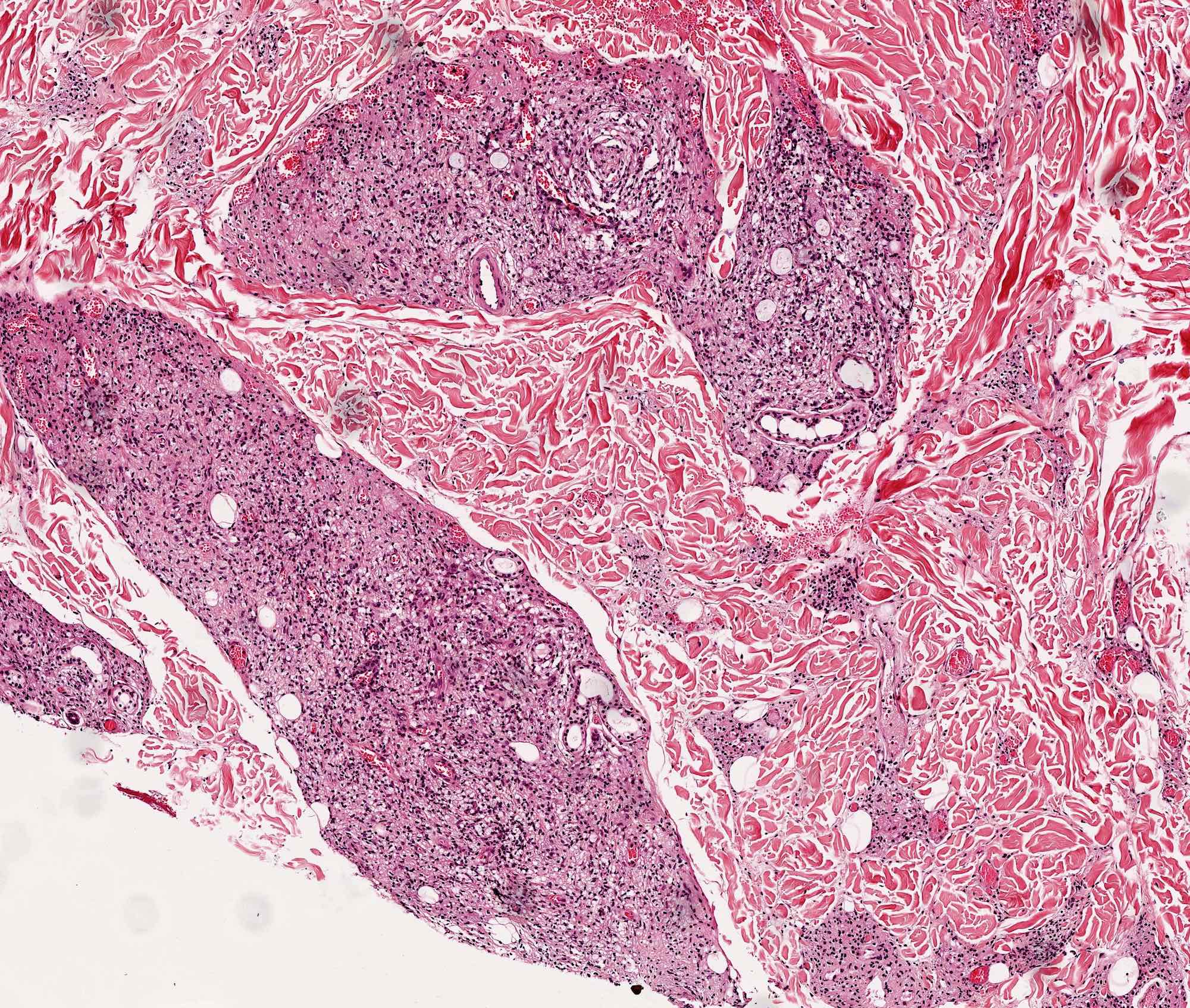
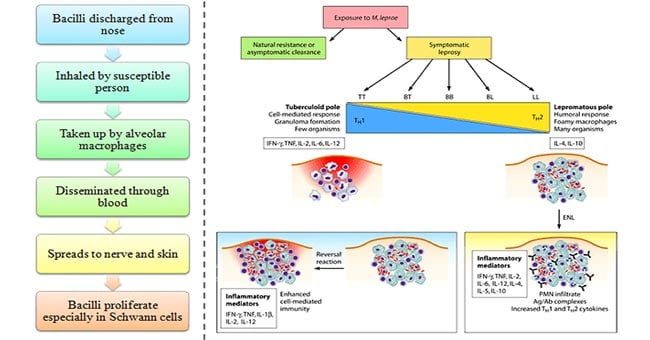
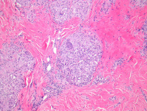
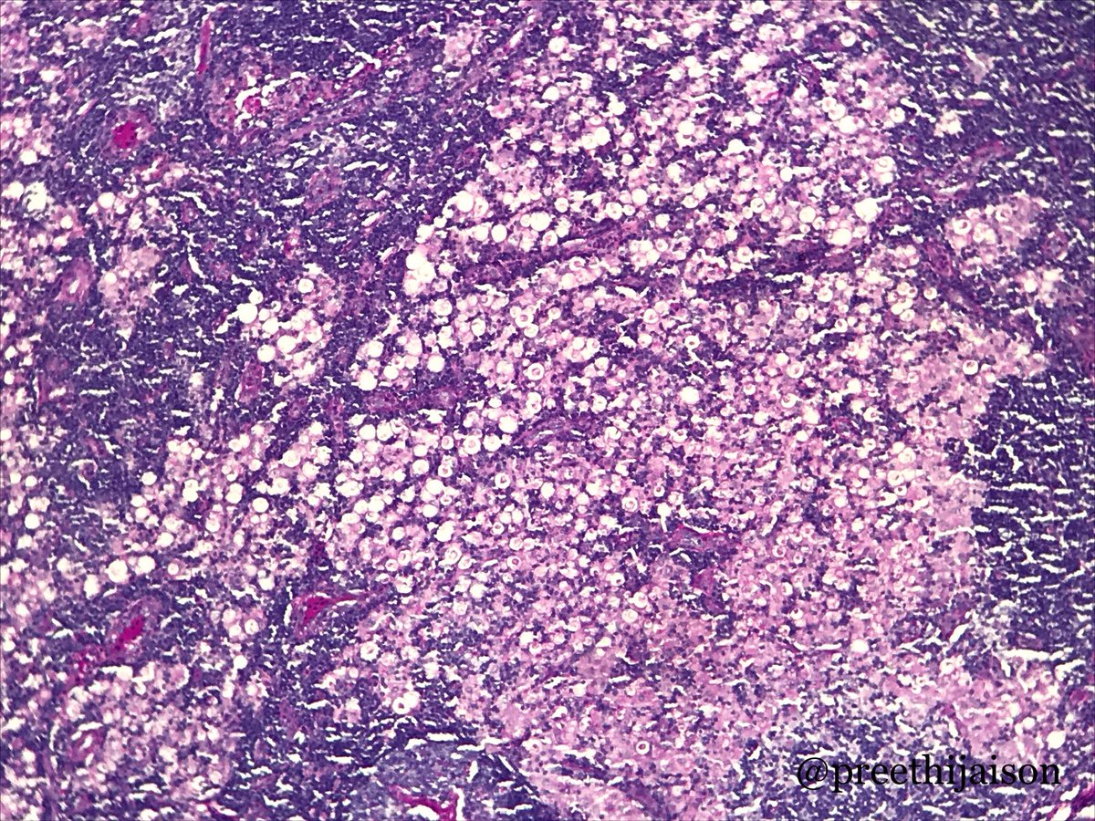
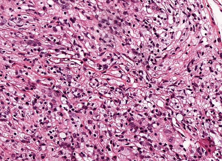
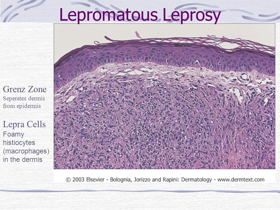
This debilitating form of leprosy begins to spread causing the eyebrows to disappear and spongy tumor like swellings appear on the face and body. Large numbers of mycobacterium leprae chiefly in masses within the lepra cells often grouped together like bundles of cigars or arranged in a palisade multibacillary diseasethe cell mediated immune cmi response to organism is poor and the lepromin test is negative. Lepromatous leprosy is characterized by a poor immunologic response and reveals diffuse infiltration by histiocytes that are incapable of undergoing epithelioid transformation and unable to form granulomas.
The granulation tissue with a large aggregation of vacuolated mononuclear cells containing lepra bacilli is a characteristic feature of lepromatous to repeated trauma and may get secondary infected resulting to distortion and mutilation of extremities. Lepromatous leprosy in contrast to the tuberculoid form of leprosy is characterized by the absence of epithelioid cells in the lesions. Bacteria are present in large numbers in cutaneous nerves and in endothelium and media of small and large vessels.
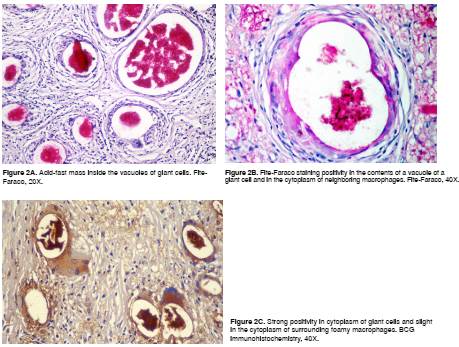
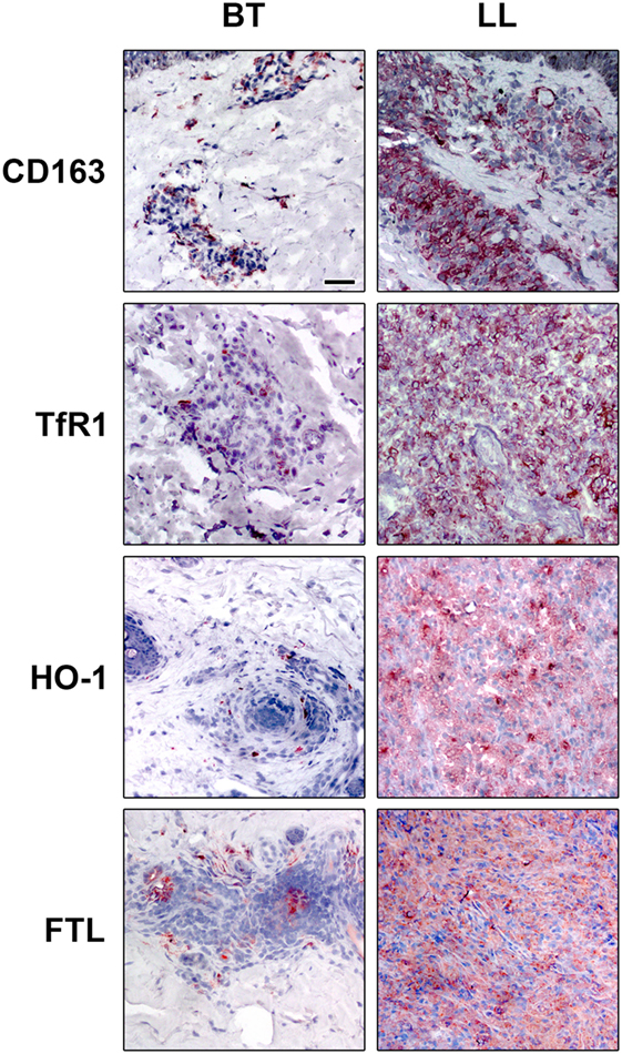
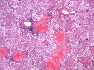
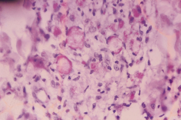
Peripheral disease predominates clinically history of residence in area where leprosy is endemic spleen may contain clusters of macrophages filled with acid fast organisms lepra cells. Distinctive large mononuclear phagocytes macrophages with a foamlike cytoplasm and also poorly staining saclike structures resulting from degeneration of such cells observed characteristically in leprous inflammatory reactions. Lepromatosis are the mycobacteria that cause leprosy.
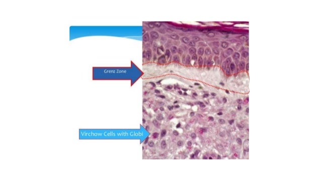
Epithelioid cells and giant cells are not found. Lepromatous leprosy involvement is most intense in skin nerves and extremities. Macrophages may be distended with large groups of leprosy bacilli globi.
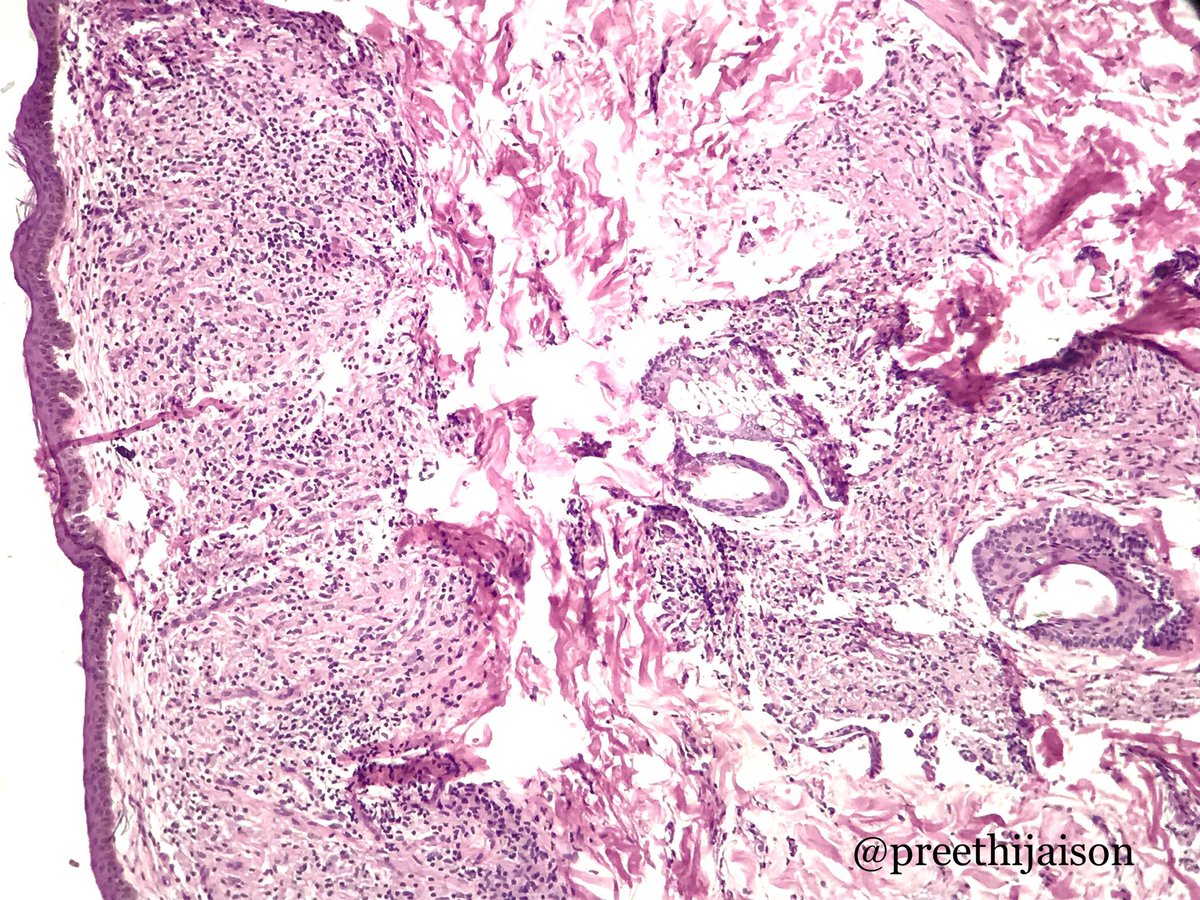
The disease attacks the internal organs bones joints and marrow of. Granulomas are most numerous around blood vessels nerves and skin appendages. In this form of leprosy mycobacterium leprae are found in lesion in large numbers.
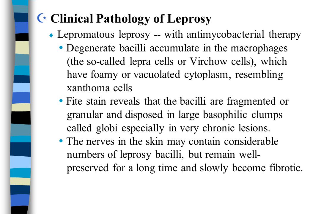
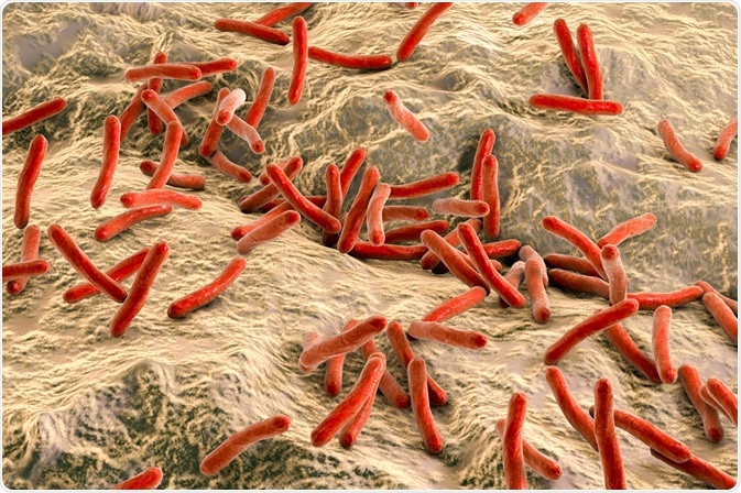
Leprae is an intracellular acid fast bacterium that is aerobic and rod shaped. Lepromatosis is indistinguishable clinically from m. These foamy histiocytes lepra cells virchow cells may be stuffed with abundant mycobacilli globi which remain uncleared by the host.

